
Proteomics-based evaluation of AAV dystrophin gene therapy outcomes in mdx skeletal muscle
Apr 24, 2026
The 2026 jci.insight by Johnson, E.E., et al. researches Proteomics-based evaluation of AAV dystrophin gene therapy outcomes in mdx skeletal muscle.
Introduction
Duchenne muscular dystrophy (DMD) is a fatal genetic muscle-wasting disease characterized by loss of dystrophin protein. Therapeutic attempts to restore a functional copy of dystrophin to striated muscle are under active development, and many utilize adeno-associated viral (AAV) vectors. However, the limited cargo capacity of AAVs precludes delivery of full-length dystrophin, a 427 kDa protein, to target tissues. Recently, we developed a method to express large dystrophin constructs using the protein trans-splicing mechanism mediated by split inteins and myotropic AAV vectors. The efficacy of this approach to restore muscle function in mdx4cv mice was previously assessed using histology, dystrophin immunolabeling, and Western blotting. Here, we expand our molecular characterization of dystrophin constructs with variable lengths using a mass spectrometry–based proteomics approach, providing insight into unique protein expression profiles in skeletal muscles of wild-type, dystrophic mdx4cv, and AAV-treated mdx4cv mice. Our data reveal several affected cellular processes in mdx4cv skeletal muscles with changes in the expression profiles of key proteins to muscle homeostasis, whereas successful expression of dystrophin constructs results in an intermediate to complete restoration. This study highlights several biomarkers that could be used in future preclinical or clinical studies to evaluate the effectiveness of therapeutic strategies.
Finding Highlights
- Described a method for delivering and expressing large dystrophins using protein trans-splicing (PTS) mediated by split inteins and myotropic AAV vectors.
- Describe a sensitive mass spectrometry–based (MS-based) proteomics workflow that allows a holistic analysis of protein expression of wild-type (WT), saline-, or AAV-treated mdx4cv mice.
- Reveal subtle changes in dystrophic muscles expressing different dystrophin-based constructs and led to the identification of cellular biomarkers with variable expression profiles.
Key Results and Figure Interpretation
- Validation of dystrophin gene therapy replacement (Fig.1 and Fig.2).
We employed an isobaric labeling multiplex discovery proteomics approach to compare the skeletal muscle proteomes of healthy (WT), dystrophic (mdx4cv), and AAVMYO1-treated mdx4cv mice with variable dystrophin constructs. Myotropic AAVMYO1 vectors were administered systemically into 8-week-old mice at low doses of 2×10¹³ vg/kg to express μDys5 (ΔSR2-15, Δ18-21, ΔCT) from a single vector or midi-Dys (ΔSR5-15) from dual vector, whereas triple AAVMYO1 were mixed and injected at a total dose of 4×10¹³ vg/kg to express full-Dys (Figure 1, A-C). Three months later, gastrocnemius muscles were collected from 6 AAV-treated mice as well as age-matched saline-treated mdx4cv and WT mice. Protein lysates were extracted and labeled with TMT isobaric tags, and 2 proteomics screens were conducted (Figure 1D).
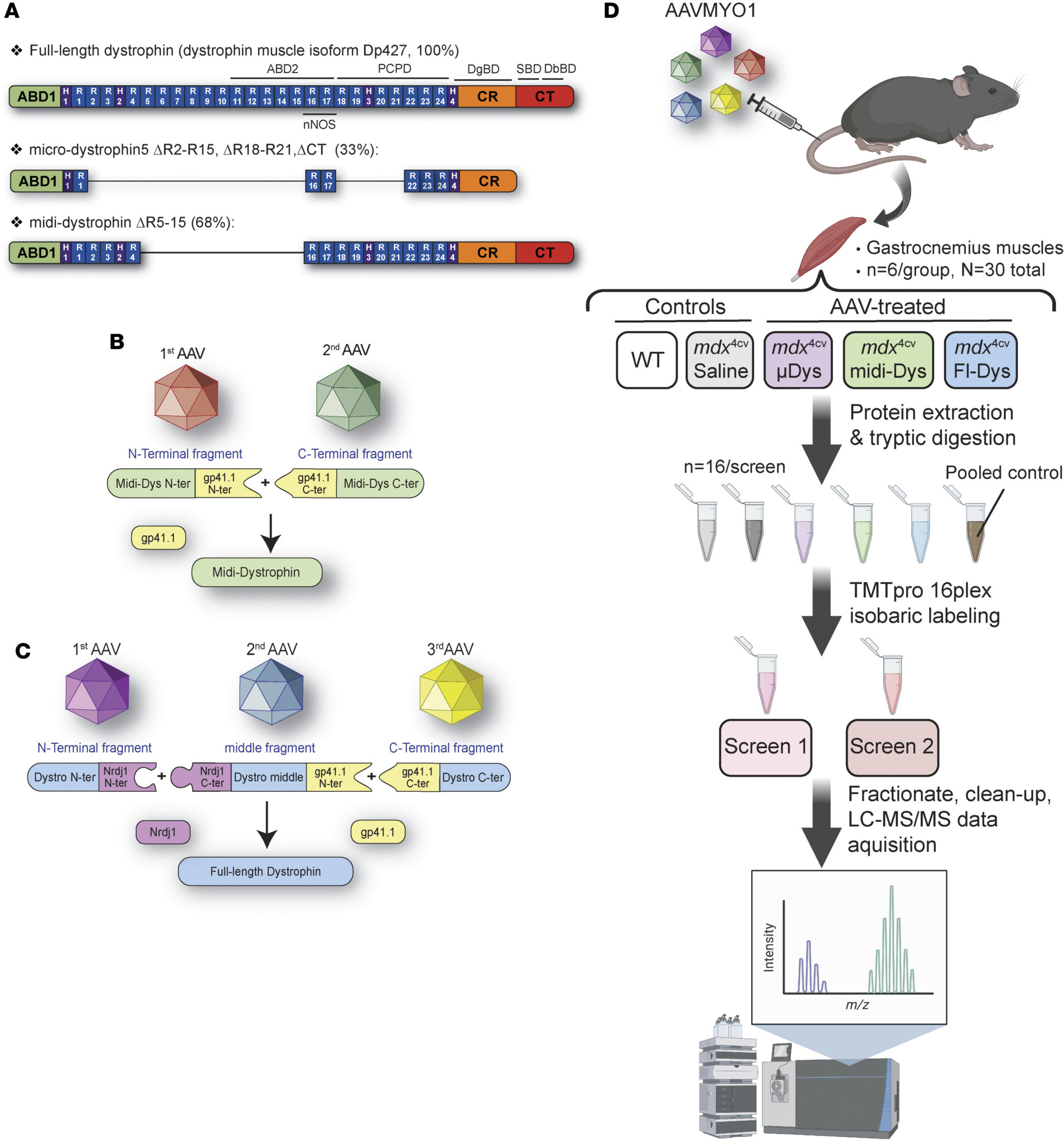
Figure 1. Schematic representation of dystrophin clones tested, split intein approach to express large constructs, and proteomics workflow.
Transgenic dystrophin constructs were detected in the samples from mdx4cv mice treated with single, dual, or triple AAVs, but at lower abundance versus endogenous dystrophin in WT muscles (Figure 2A). As expected, the average abundance of peptide sequences specific to fullDys was found elevated exclusively in WT or triple-AAV groups (Figure 2B). Similarly, peptide sequences specific to transgenic/human dystrophins (μDys5, intein-generated midi-Dys, or full-Dys) were elevated across all AAV treatment groups (Figure 2C). Using different peptides, μDys and midi-Dys were detected at comparable levels, whereas average full-Dys abundance was slightly lower. Finally, by searching peptides specific to large dystrophin (i.e., excluding μDys), we confirmed the exclusive expression of large dystrophins in WT or mdx4cv mice treated with dual or triple AAV vectors (Figure 2D). Overall, the abundance of the dystrophins was peptide dependent, with a variable sensitivity observed from one peptide to another.
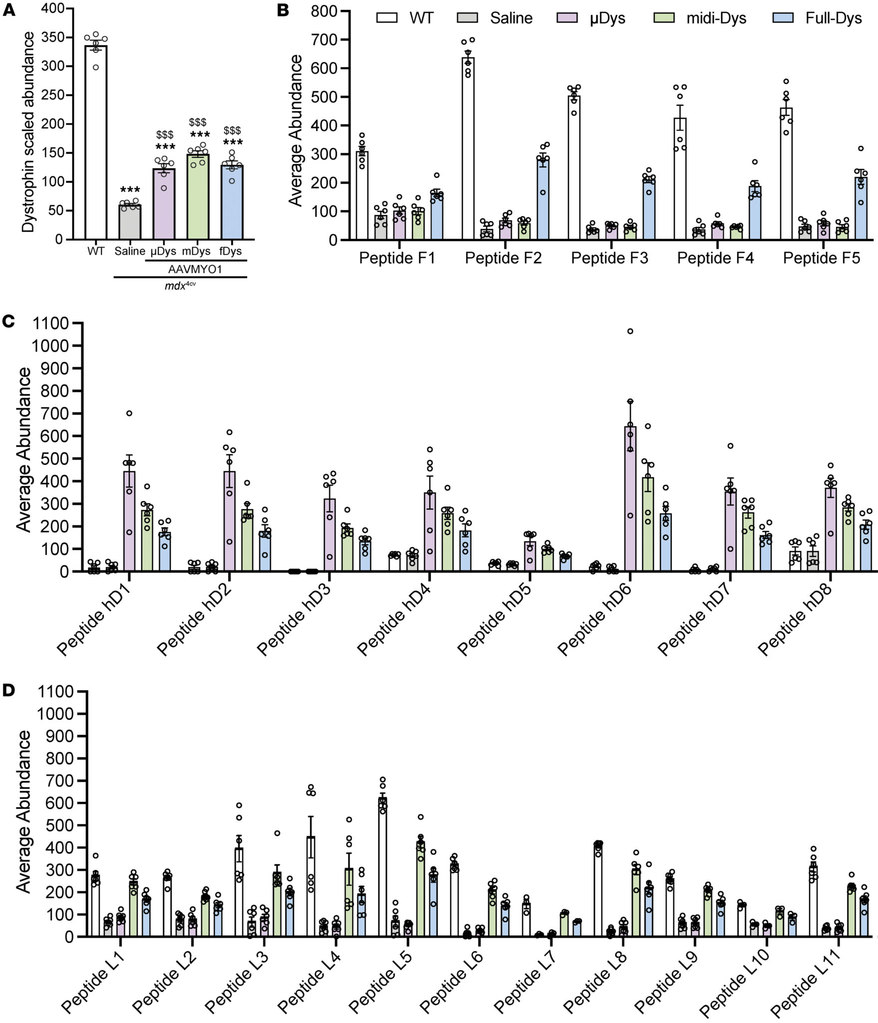
Figure 2. Detection of dystrophin expression and quantification of peptide-specific abundance using proteomics
- Muscle histology improvement after dystrophin expression (Fig.3).
To evaluate the muscle histology and compare the therapeutic benefits of each gene replacement modality, serial cross sections of gastrocnemius were stained with hematoxylin and eosin (H&E), trichrome, or immunolabeled using specific antibodies raised against elements of the DGC or periostin. In the group treated with AAVs, dystrophin expression was detected in 40%–60% of myofibers, whereas a few revertant fibers, not exceeding 1%, were found in the saline group (Figure 3, A and B). Muscles from animals treated with saline presented typical dystrophic muscle histology with small fibers and fibrotic and infiltrated muscle tissue compared with WT muscles (Figure 3, A, and C-E). A marked reduction in collagen content was also found in groups treated with AAVs (Figure 3, A and C). Similarly, immunolabeling of periostin showed an increased area in saline-treated dystrophic muscles, confirming the expansion of the ECM, which AAV-dystrophin treatment prevented (Figure 3F). Interestingly, while proteomics data confirmed the upregulation of periostin (Figure 3G), variable abundance of collagen isoforms was observed. For instance, collagen isoform I (α1 and α2), IV (α1 and α2), VI (α1, α2, and α3), and XII abundance was unchanged among groups, whereas collagen type III (α1), V (α2 and α3), VI (α6), and XIV (α1 chain) abundance was elevated in mdx4cv muscle and restored by AAV treatment (Figure 3H).
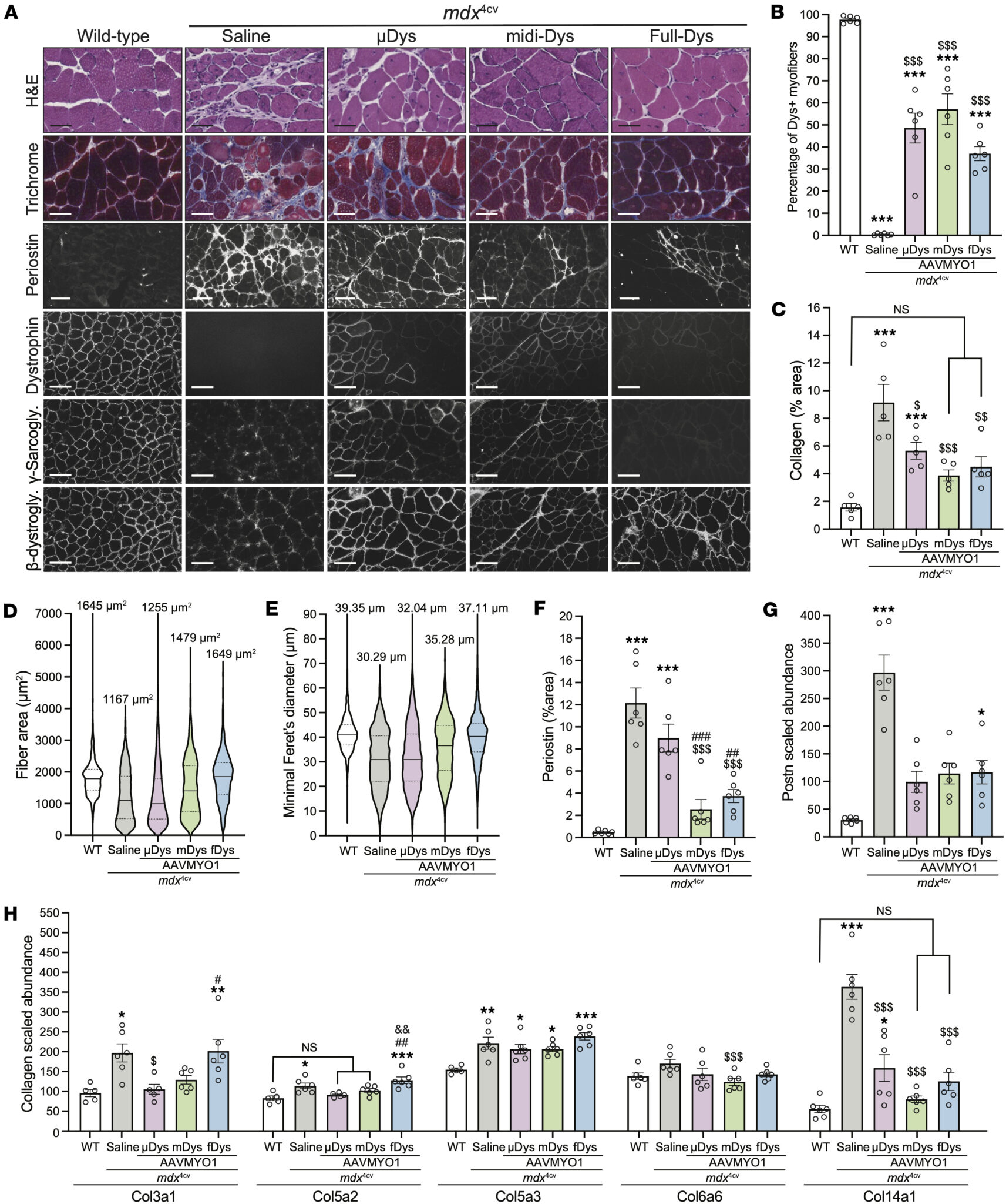
Figure 3. Histology analysis of gastrocnemius muscle cross sections showing improvements with dystrophin constructs
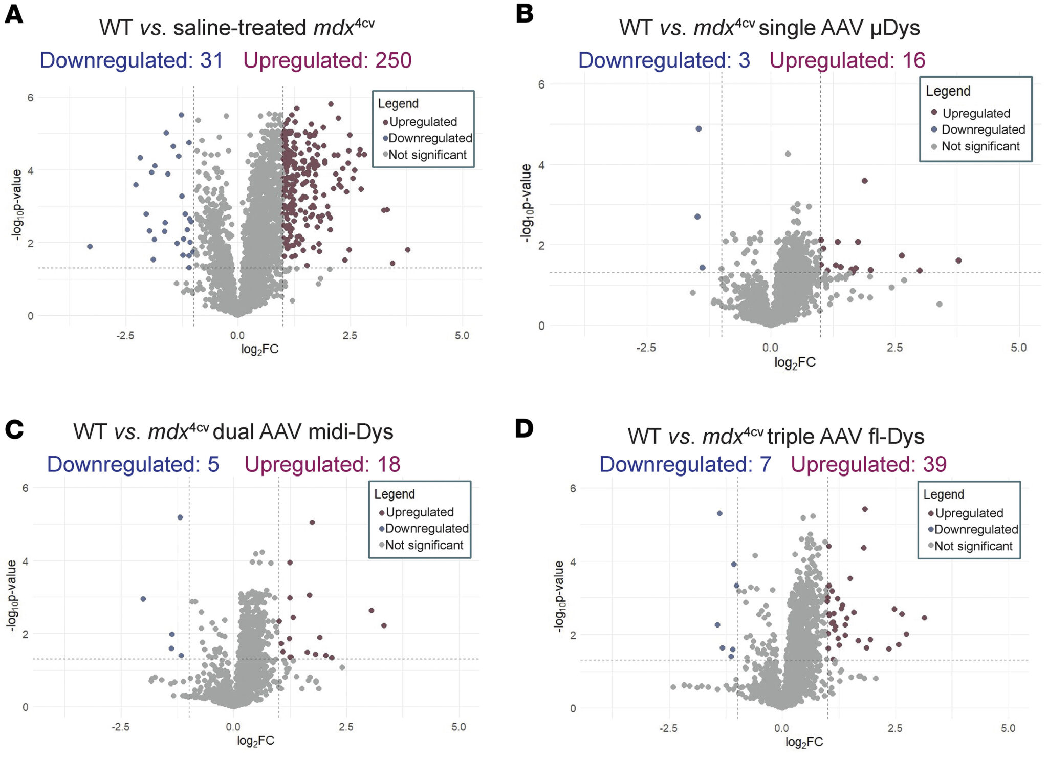
Figure 4. Comparison of protein expression profiles between experimental groups
- Characterization of molecular changes using proteomics (Fig.4, Fig.5 and Fig.6).
Dystrophin-deficient mdx4cv gastrocnemius muscle displayed a large number of differentially expressed proteins (DEPs) compared with WT muscle, including 250 upregulated proteins and 31 downregulated proteins (Figure 4A). The top upregulated and downregulated pathways in mdx4cv muscle have been previously reported in mdx mice, demonstrating defects in, for example, cytoskeletal structure and sarcolemmal integrity, ECM organization, and fatty acid metabolism. In contrast, few proteins displayed significantly elevated or depleted levels in single, dual, or triple AAV–treated mdx4cv gastrocnemius muscle compared with WT muscle (Figure 4, B–D). A total of 16 upregulated and 3 downregulated proteins were identified between μDys5-mdx4cv and WT mice (Figure 4B). Eighteen upregulated proteins and 5 downregulated proteins were observed in midi-Dys-mdx muscle compared with WT (Figure 4C), while 39 upregulated and 7 downregulated proteins were found between full-Dys-mdx4cv and WT mice (Figure 4D).
Further analysis of top upregulated and downregulated DEPs in mdx4cv compared with WT gastrocnemius revealed that several cellular processes were dysregulated (Figure 5). For instance, upregulated DEPs in mdx4cv muscle were enriched for molecular functions and biological processes, including protein and mRNA binding, cytoskeletal structure, supramolecular fiber organization, and regulation of RNA splicing, with cellular compartment enrichment for cytoplasmic, collagen-containing ECM, spliceosome, sarcolemmal, and endoplasmic reticulum proteins (Figure 5, A and B). Downregulated DEPs in mdx4cv muscles, however, were enriched for molecular functions including nucleosome DNA binding, fatty acid metabolic processes, and muscle tissue development, with the cellular compartment enrichment for DGC, sarcolemmal, cytoplasmic, and euchromatin-enriched proteins (Figure 5, A and C).
Importantly, several of these defects were partially restored with the dystrophin replacement using AAVMYO1 vectors at variable levels (Figures 5 and 6). For example, treatment with AAV-μDys5 restored the abundance of DGC proteins, In contrast, levels of α-syntrophin and utrophin were normalized with dual AAV-midi-Dys but remain slightly affected with μDys or triple AAV-full-Dys treatments (Figure 6, C and D). Similarly, several proteins with elevated abundance in WT muscle displayed reduced abundance in saline-treated mdx4cv, whereas dystrophin construct expression mediated by AAV partially or fully restored their cellular enrichment, including protein-arginine deiminase type-2 and myoglobin (Figure 6E). In contrast, tubulin β6 class V, whose abundance was higher in the saline group, consistent with a previous study was greatly reduced in AAV-treated groups (Figure 6E)
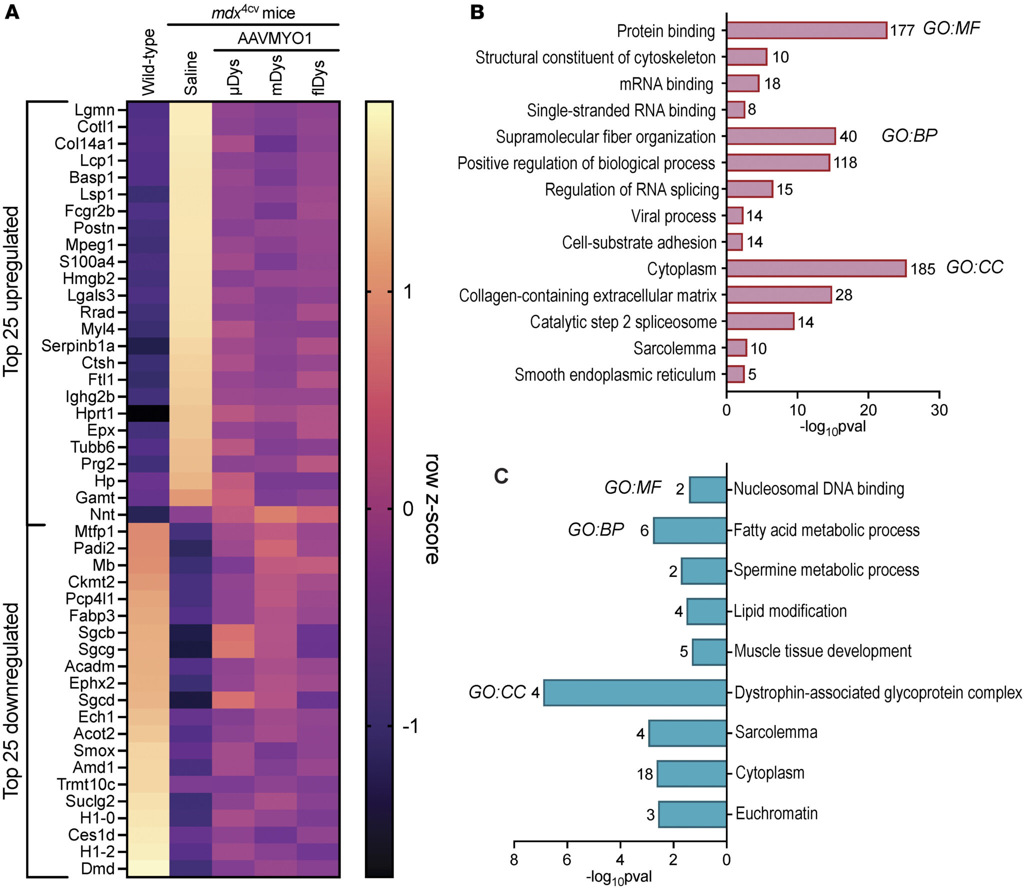
Figure 5. Analysis of protein expression profile demonstrates proteomic rescue by dystrophin constructs.
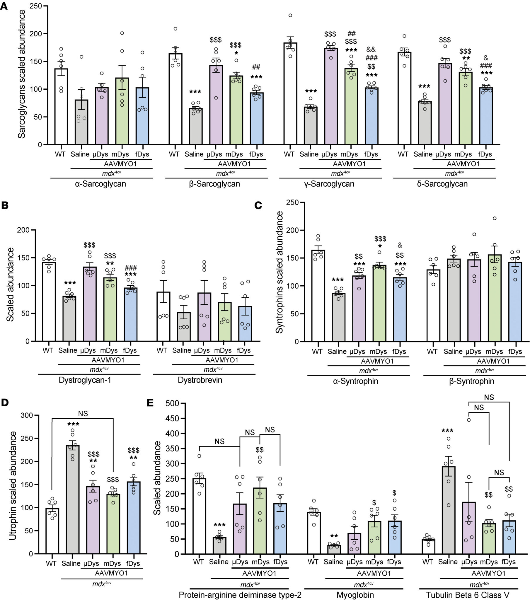
Figure 6. Dystrophin delivery alleviates DGC protein defects in mdx mice
- Dystrophin replacement partially restores biomarkers involved in membrane repair and myogenesis (Fig.7)
A general trend of pathway elevation was observed in mdx4cv muscle, with partial restoration across all AAV-Dys treatment groups (Figure 7A). Following this pattern, annexin A1 and annexin A5 levels were increased in mdx4cv muscle compared with WT and partially restored by AAV-Dys treatment (Figure 7B). We also observed elevated annexin A4 levels in mdx4cv muscle, but only AAV-midiDys treatment significantly reduced annexin A4 to an intermediate level between WT and dystrophin-deficient muscle (Figure 7B). Dysferlin also displayed elevated levels in saline-mdx4cv muscles. While an intermediate restoration was detected in the AAV-treated groups, only dual midi-Dys treatment significantly reduced dysferlin levels compared to saline-mdx4cv (Figure 7C). Likewise, elevated levels of caveolin-3 and MG53/TRIM72 were found in control mdx4cv muscle that were significantly but modestly reduced by AAV treatment (Figure 7C).
Furthermore, we analyzed the expression level of proteins implicated in membrane remodeling, trafficking, and cytoskeleton dynamics, such as clathrin light chain A, dynamin-2, and amphiphysin-2 (BIN1). These proteins were enriched in saline-mdx4cv muscles, with 2- to 3-fold higher levels compared with WT muscles (Figure 7D). However, variable effects were found with the different dystrophin constructs. For instance, partial restoration was observed with the single AAV-μDys treatment, whereas near-complete normalization of these proteins was obtained with dual or triple AAV approaches (Figure 7D). Conversely, all dystrophin constructs restored the level of galectin-1 to WT levels and significantly reduced galectin-3, which were found to be 3- and 5-fold higher, respectively, in saline-treated dystrophic muscles (Figure 7, E and F).
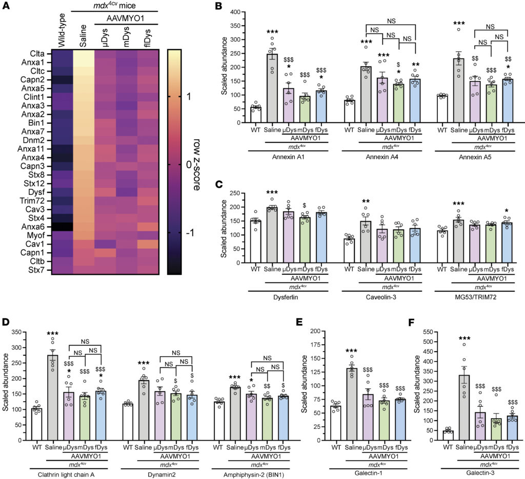
Figure 7. Amelioration of altered membrane repair and myogenesis pathway markers in mdx4cv muscle mediated by AAV-dystrophin constructs
- Incomplete corrections with dystrophin gene therapy (Fig.8).
We filtered our dataset for proteins that met the following 2 criteria: (a) significantly altered in AAV-treated mdx4cv muscle compared with WT muscle, and (b) not significantly altered between AAV-treated and untreated mdx4cv groups.
After filtering, we obtained short lists of unrestored DEPs in μDys5-mdx4cv, midi-Dys-mdx4cv, and full-Dys-mdx4cv gastrocnemius muscle (Figure 8, A–C). Several proteins demonstrated depleted abundance in mdx4cv muscle that was not restored by the different dystrophin constructs, including carboxylesterase 1D (gene name Ces1d; Figure 8D), spermine oxidase (gene name Smox; Figure 8E), tRNA methyltransferase 10 homolog C (gene name Trmt10c; Figure 8F), adenosylmethionine decarboxylase (gene name Amd1; Figure 8G), and histone H1.2 (gene name H1-2; Figure 8H). Levels of several upregulated proteins in mdx4cv muscle were not ameliorated or were only partially ameliorated by AAV-dystrophin treatments, including myosin light chain 6B (gene name Myl6b; Figure 8I) and heme-binding protein 1 (gene name Hebp1; Figure 8J). Importantly, the introduction of split-intein dystrophin constructs did not induce unique or deleterious proteomic changes in the mdx4cv gastrocnemius muscles. A singular protein, nicotinamide nucleotide transhydrogenase (NNT; gene name Nnt), demonstrated expression changes in mdx4cv muscle that were more pronounced with AAV-dystrophin treatment; however, NNT expression levels did not display a statistically significant difference between treated and untreated mdx4cv muscle (Figure 8K). Only 2 of the proteins identified as dysregulated in naive or AAV-treated mdx4cv muscle, myosin light chain 4 (gene name Myl4) and hypoxanthine-guanine phosphoribosyl transferase (gene name Hprt1), were referenced in previous studies involving mdx mice. Notably, a singular protein, eukaryotic translation initiation factor 2D (gene name Eif2d), was identified as uniquely altered by AAV treatment (Figure 8L), suggesting a minimal biological impact of injection with the AAV constructs themselves.
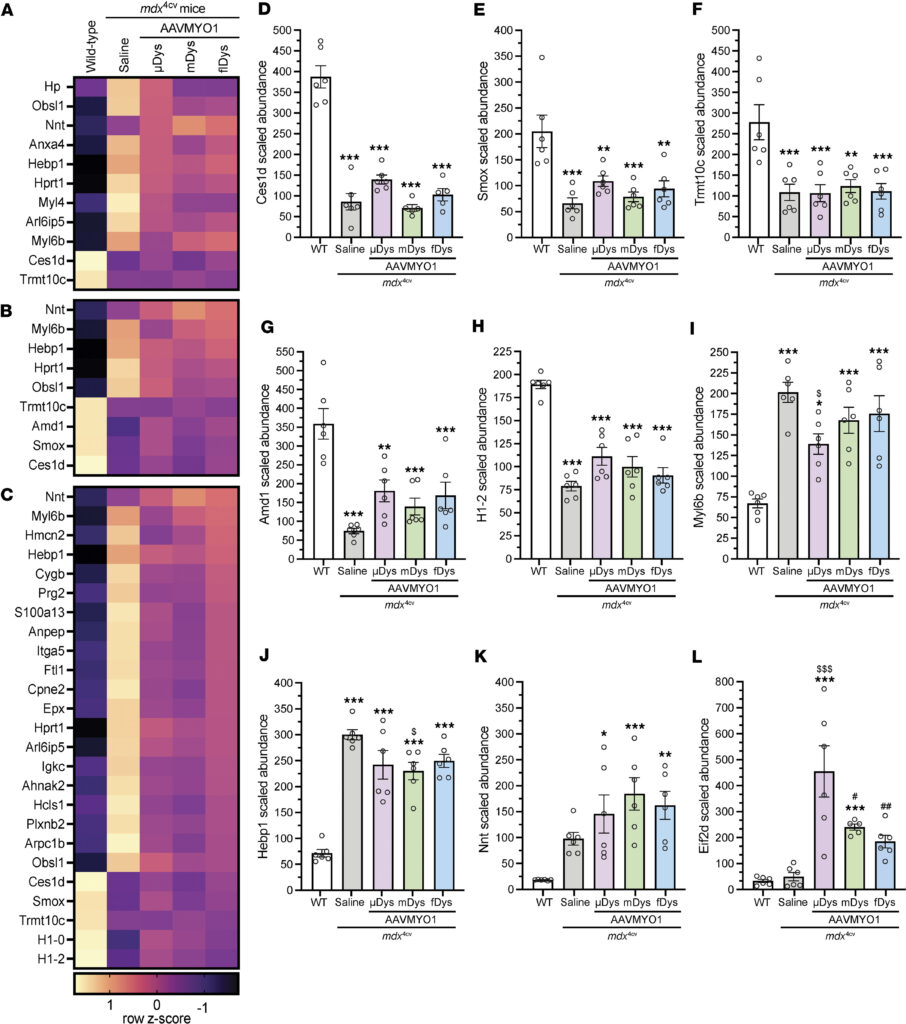
Figure 8. Proteins with unrestored expression in mdx4cv mice treated with various dystrophin constructs.
Conclusion
Our dataset confirms the depletion of DGC proteins and demonstrates an overall pattern of elevated expression for membrane trafficking and repair pathway proteins in mdx4cv muscle, with partial restoration of some proteins by the different dystrophin constructs expressed via AAV delivery. Lower levels of DGC proteins were previously described in dystrophin-null muscles as a direct consequence of the absence of dystrophin, whereas the upregulation of MG53/TRIM72, dysferlin, caveolin-3, utrophin, and members of the annexin family may reflect an adaptive response to increased sarcolemmal membrane fragility and rupture.
The function of annexin A4 in sarcolemmal repair has not been clearly defined. However, overexpression of other annexin family members has been observed in DMD muscle as a response to increased membrane fragility and activation of membrane repair processes. Interestingly, genetic mutations affecting the expression of dysferlin and caveolin- have been associated with the development of muscular dystrophies.
In conclusion, this study describes the use of a proteomics approach to study the global protein expression in healthy or dystrophic skeletal muscles. This method can be implemented to validate therapeutic strategies in preclinical and clinical studies and monitor the effectiveness of treatments for muscular disease.
Reference
Johnson, E.E., et al., Proteomics-based evaluation of AAV dystrophin gene therapy outcomes in mdx skeletal muscle. JCI Insight, 2026. 11(2).