
Safety and Efficacy of cceAAV-aflibercept-Fc-YTE (F-CRG-B191): A Gene Therapy for Neovascular Age-Related Macular Degeneration
May 15, 2026
The 2026 Invest Ophthalmol Vis Sci by Shi, J., et al. researches Safety and efficacy of cceAAV-aflibercept-Fc-YTE (F-CRG-B191): A gene therapy for neovascular age-related macular degeneration.
Introduction
To evaluate the safety and efficacy of a novel gene therapy vector, covalently closed-end double-stranded adeno-associated virus-aflibercept-Fc-YTE (cceAAV-aflibercept-Fc-YTE; clinical trial name F-CRG-B191), designed for sustained anti-vascular endothelial growth factor (VEGF) expression in the treatment of neovascular age-related macular degeneration (nAMD). Compared to conventional AAV vectors expressing aflibercept, cceAAV-aflibercept-Fc-YTE significantly reduced the incidence of grade IV CNV lesions in both mice and NHP models. In the human subject, F-CRG-B191 administration led to a sustained reduction in CNV area and improvement in visual acuity. No significant immune response or retinal toxicity was observed in either preclinical models or the treated patient.
Finding Highlights
- Developed cceAAV-aflibercept-Fc-YTE (clinical trial name F-CRG-B191), a cceAAV-based gene therapy drug designed to treat nAMD by expressing aflibercept to inhibit VEGF.
- Evaluate the preclinically for safety and efficacy of cceAAV-aflibercept-Fc-YTE.
- In the clinical study, FCRG-B191 achieved anticipated therapeutic effects without severe adverse effects.
Key Results and Figure Interpretation
- Expression of cceAAV-Affibercept-Fc-YTE in Mice (Fig.1).
cceAAV-aflibercept-Fc-YTE was constructed using cceAAV carrying an improved gene encoding aflibercept-Fc-YTE, a midriffed format of the VEGF antibody (Fig. 1A). The key elements of cceAAV-aflibercept-Fc-YTE are illustrated in Figure 1, and the schematic structures of three different AAVs including cceAAV, scAAV, and ssAAV are highlighted in Figure 1B. The cceAAV-affibercept-Fc-YTE was delivered into the eyes of the C57BL/6J mice via subretinal injection. The expression of aflibercept-Fc-YTE protein showed a dose-dependent increase, with 69.45±17.19 ng/eye in the 3 x10⁷ GC dosing group (n = 4), 326.75 ±59.96 ng/eye in the 1×10⁸ GC dosing group (n = 4), and 1644.10±288.43ng/eye in the 6x 10⁸ GC dosing group (n = 4). To assess the transgene efficiency, aflibercept-Fc-YTE expression was compared among cceAAV, scAAV, and ssAAV vectors. At a dose of 3×10⁷ GC/eye, aflibercept-Fc-YTE expression in the cceAAV group was 1.9 times higher than in the scAAVgroup (P = 0.0493). As the dose increased, the advantage ofcceAAV also increased. At a dose of 1×10⁸ GC/eye, protein expression in the cceAAV group was 2.5 and 3.2 times higherthan in the ssAAV and scAAV groups, respectively (all P <0.001). At a dose of 6 x10⁸ GC/eye, protein expression inthe cceAAV group was 3.5 and 4.9 times higher than in the AAV and scAAV groups, respectively (Fig.1C). These results indicate that this novel cceAAV vector enhanced aflibercept expression in vivo. The expression of aflibercept-Fc-YTE in the eyes of mice (6.7×10⁸ GC/eye) was 906.05±572.38 ng/eye at week 2 and reached a peak of 2332.81±333.39 ng/eye at week 4. Expression gradually decreased and stabilized at approximately 1186.25±140.63ng/eye after week 8, persisting until the final observation at week 24 (Fig. 1D).
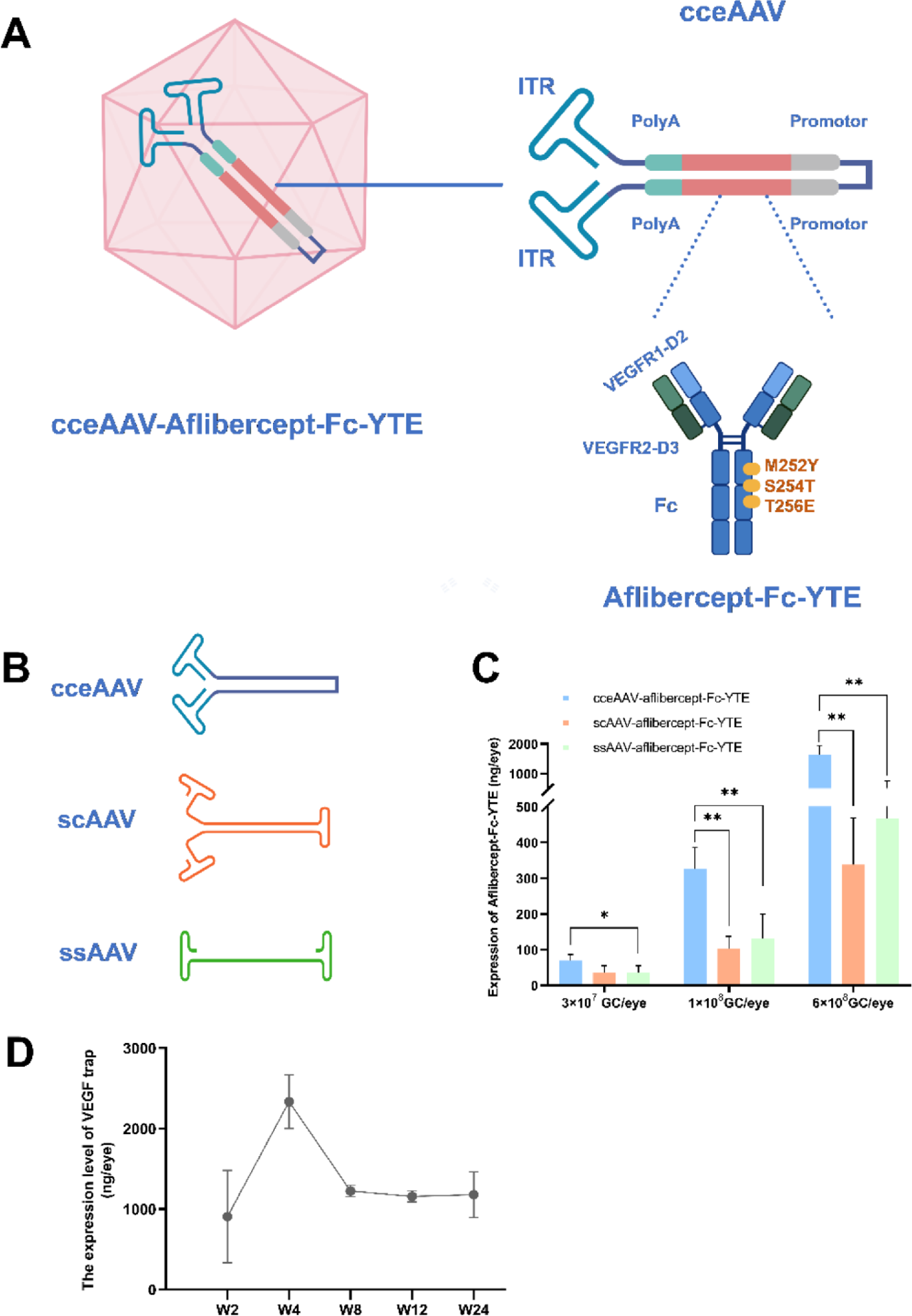
Figure 1. Structure of cceAAV-aflibercept-Fc-YTE and transgene expression comparison among different vectors.
- Efficacy of Aflibercept-Fc-YTE in Mice CNV Model (Fig.2).
The evaluation timeline of cceAAV treatments is illustrated in Figure 2A. Vehicle (formulation buffer, as a negative control) or cceAAV-aflibercept-Fc-YTE and cceAAV- aflibercept at different dosages were injected into the subretinal space of C57BL/6J mice (week 0). Four weeks later, retinal laser photocoagulation was performed to induce CNV, similar with the previous study. Compared to the vehicle group, the CNV area was significantly reduced in all cceAAV-aflibercept-Fc-YTE groups (all P < 0.001). The highest dose yielded the best results (3 x10⁷ GC/eye vs. 1 x10⁹ GC/eye, P = 0.004; 1 x10⁸ GC/eye vs. 1×10⁹ GC/eye, P = 0.011). Compared to the vehicle group, the CNV area was also significantly reduced in all cceAAV- aflibercept groups (all P < 0.001). At the same dose of 1×10⁹ GC/eye, the CNV area in the cceAAV-aflibercept-Fc-YTE group was significantly smaller than in the cceAAV-aflibercept group (P = 0.0015) (Fig. 2B). Compared to the vehicle group, the CNV lesion grading was significantly reduced in the cceAAV-aflibercept-Fc-YTE, cceAAV-aflibercept, and positive control groups (all P < 0.001). There were no differences in the CNV lesion grading among the three different doses of cceAAV-aflibercept-Fc-YTE or cceAAV-aflibercept. The CNV lesion grading of the cceAAV-aflibercept-Fc-YTE group (1 × 10⁹ GC/eye) was significantly lower than that of the cceAAV-aflibercept group (1 × 10⁹ GC/eye; P = 0.0029) (Fig. 2C). These results indicate that the modified aflibercept-Fc-YTE shows comparable efficacy to conventional aflibercept and potentially better efficacy than cceAAV-aflibercept at the same dose (1×10⁹ GC/eye).
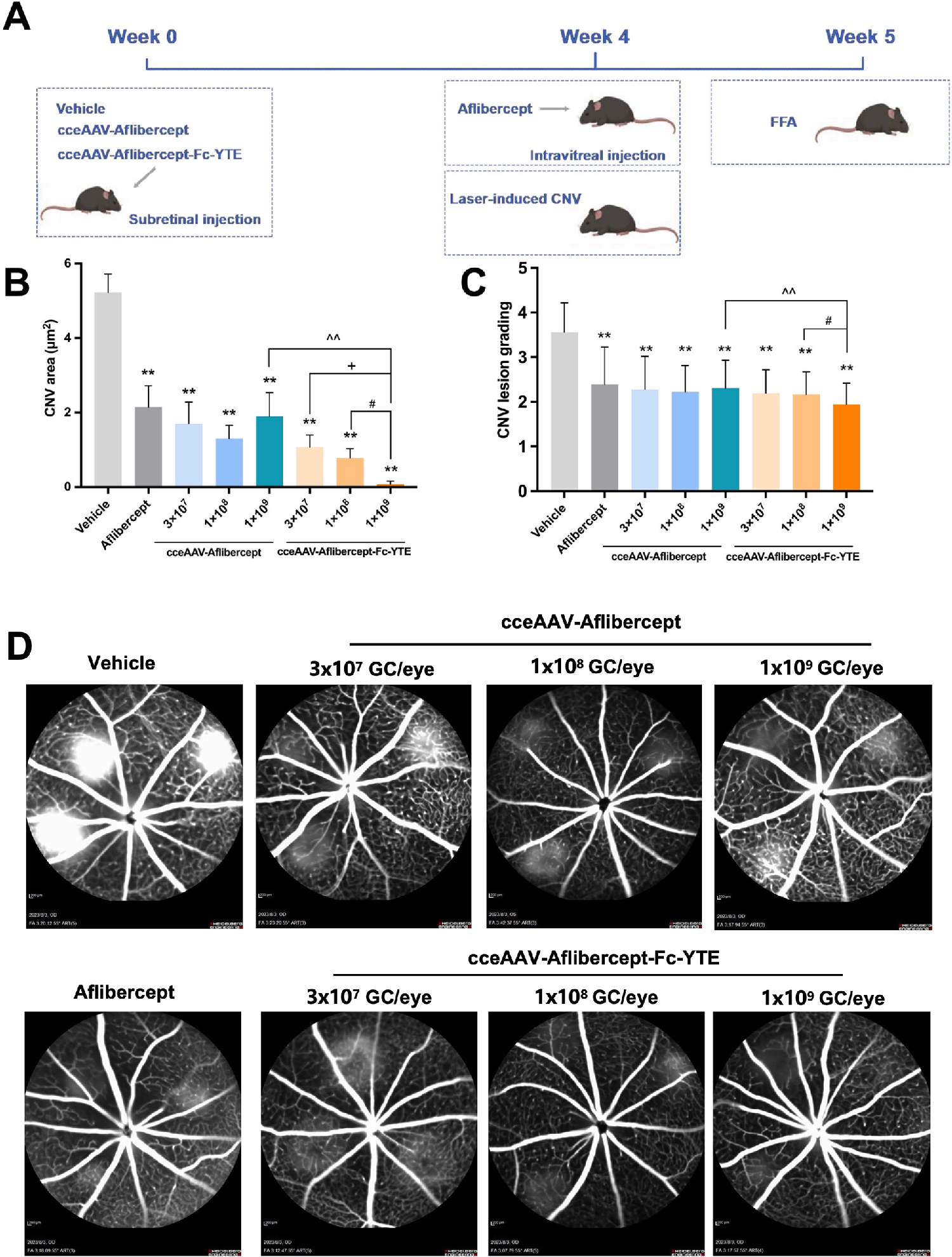
Figure 2. Efficacy of aflibercept-Fc-YTE in mice CNV model
- Retinal Structure and ERG Outcomes After Injection of cceAAV-Aflibercept-Fc-YTE in NHPs (Fig.3).
Similar to the AAV vector DNA, NAb levels were higher in the high-dose group (5 × 10¹⁰ GC/eye). In both dose groups, the levels of NAbs increased by week 2, decreased by week 4, and returned to near pre-injection levels by week 26 (Fig. 3B). These findings suggest that cceAAV-aflibercept-Fc-YTE administration did not elicit an intense immunogenic response in NHPs. OCT revealed granular hyper-reflective deposits above the RPE layer in the same region immediately after injection (Fig. 3C). The outer retinal thickness in the cceAAV-aflibercept-Fc-YTE 5 × 10¹⁰ GC/eye group significantly decreased (pre-injection: 101±0.72 μm; post-injection: 87.11 ± 2.01μm), mainly due to RPE layer thinning (pre-injection: 54.11±3.97μm; postinjection: 43.56 ± 2.94 μm). Meanwhile, no significant change was observed in the vehicle group (pre-injection: 92 ± 5.62 μm; post-injection: 92.33 ± 9.94 μm; P = 0.937) or the cceAAV-aflibercept-FcYTE 1 × 10¹⁰ GC/eye group (pre-injection: 89.33 ± 1.19 μm; post-injection: 79.56 ± 6.68 μm; P = 0.204) (Fig. 3D). Twelve weeks after the injection, scotopic ERG was performed. The amplitudes of the A-waves and B-waves (measured with dark-adapted light intensity of 0.01, 3.0, or 10.0 cd s/m2) showed no deterioration in either the cceAAV-aflibercept-Fc-YTE 1 × 10¹⁰ GC/eye group or the 5 × 10¹⁰ GC/eye group compared to the vehicle group (all P < 0.05) (Fig 3E, 3F).
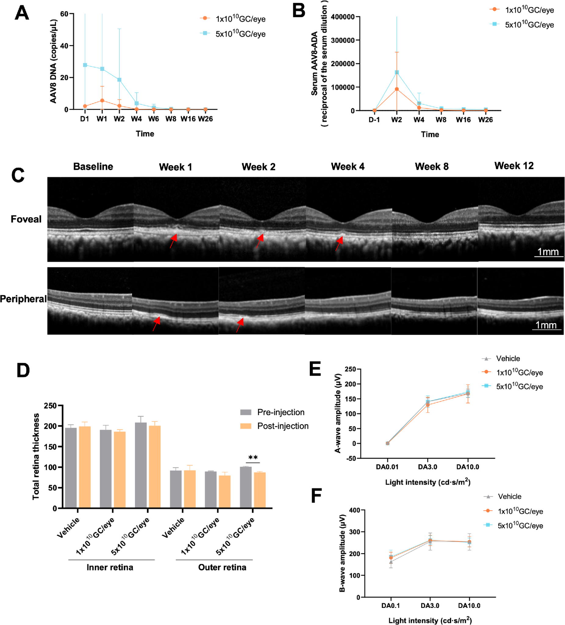
Figure 3. Safety evaluation following subretinal injection of cceAAV-aflibercept-Fc-YTE in nonhuman primates
- Efficacy Assessment of cceAAV-Aflibercept-Fc-YTE in Laser-Induced CNV Model of cceAAV-Aflibercept-Fc-YTE in NHPs (Fig.4).
The efficacy of cceAAV-aflibercept-Fc-YTE was evaluated using a laser-induced CNV model. Cynomolgus monkeys received subretinal injections of vehicle or cceAAV-aflibercept-Fc-YTE (1 × 10¹⁰ GC/eye and 5 × 10¹⁰ GC/eye). Four weeks later, the perimacular area was treated with laser photocoagulation to induce CNV. In the aflibercept group, aflibercept was injected intravitreally on the same day after laser treatment. The number of grade IV CNV lesions was determined 4 weeks after laser treatment using FFA (Fig. 4A). In the vehicle group, 62.5% of lesions were grade IV. The percentage of grade IV lesions was significantly reduced in both the cceAAV-aflibercept-Fc-YTE 1 × 10¹⁰ GC/eye group (2.1%, P = 0.0057) and the 5 × 10¹⁰ GC/eye group (4.2%, P = 0.0233). In the aflibercept group (2 mg/eye), 3.1% of lesions were grade IV. There was no significant difference among the two cceAAV-aflibercept-Fc-YTE groups and the aflibercept group (P < 0.05) (Fig. 4B).
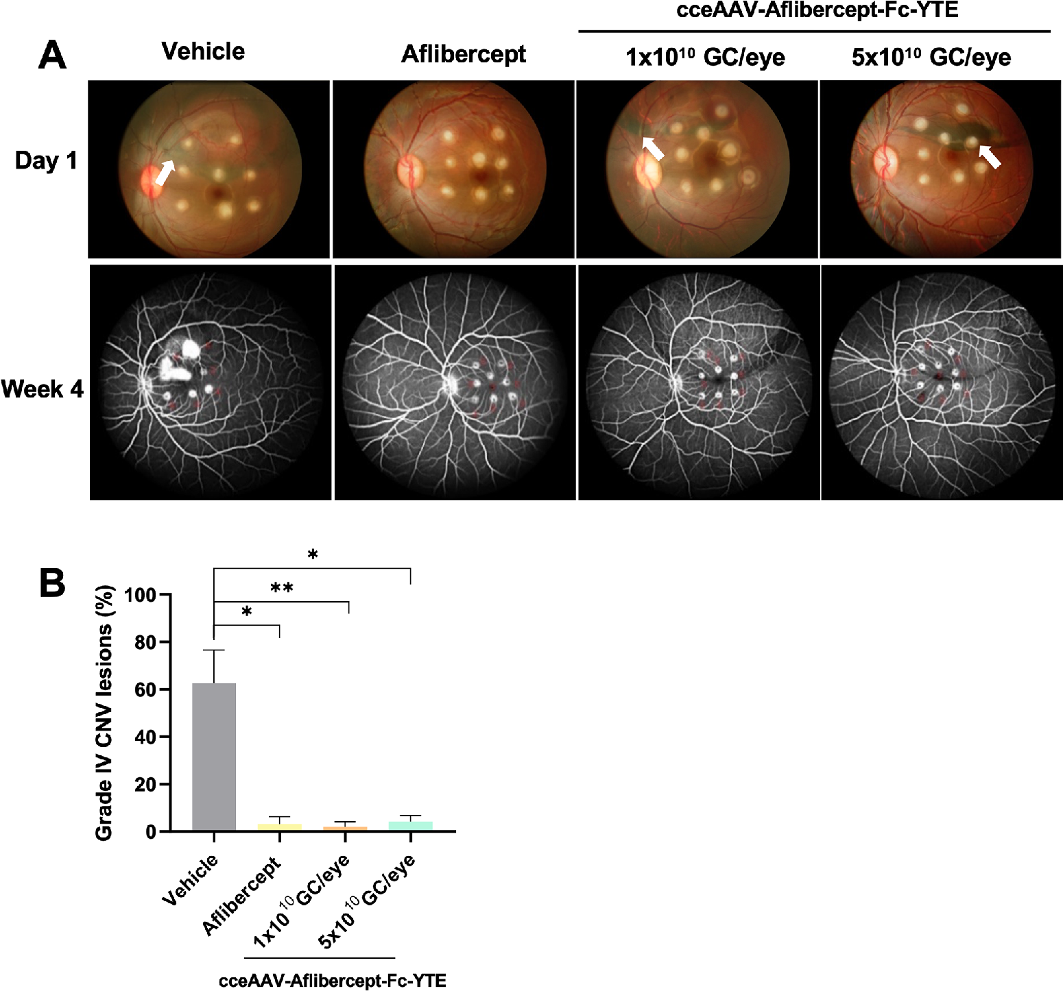
Figure 4. Efficacy evaluation of cceAAV-aflibercept-Fc-YTE in nonhuman primate eyes
- Efficacy Assessment of cceAAV-Aflibercept-Fc-YTE in Laser-Induced CNV Model of cceAAV-Aflibercept-Fc-YTE in NHPs (Fig.5 and Fig.6).
In the clinical case, the enrolled patient had received a total of 13 intravitreal anti-VEGF injections with a favorable response. Throughout the 24-week follow-up period, the patient exhibited stable IOP (Fig. 5A), and both clinical imaging and UWFP showed no significant adverse events, such as retinal hemorrhage, detachment, or abnormal pigmentation in the injection area or other areas of the fundus. However, a small area of RPE change was observed in the UWFP at the injection site 1 week after injection (Fig. 5B), with an abnormal punctate hypoautofluorescence signal on FAF. In the injected bleb region, a mottled, heterogeneous autofluorescence signal persisted throughout the follow-up period (Fig. 5C). OCT revealed no significant structural abnormalities (Fig. 5D). The patient had a baseline BCVA of 23 ETDRS letters. After injection, BCVA improved to 31 letters by 12 weeks and 44 letters by 24 weeks, representing a remarkable gain of 21 letters compared to baseline (Fig. 6A). In the macular region, no cystoid retinal edema or subretinal fluid was observed on OCT during the follow-up period. By week 24, the CRT decreased from 461 μm at base line to 365 μm. The CNV lesion area decreased from 0.41 mm2 at baseline to 0.25 mm2 (Figs. 6B–D). Throughout the follow-up period, no grade 3 or 4 common terminology criteria for adverse events (CTCAE) were reported. In addition, no injection-related adverse events occurred, further confirming the safety of F-CRG-B191 (cceAAV-aflibercept-Fc-YTE).
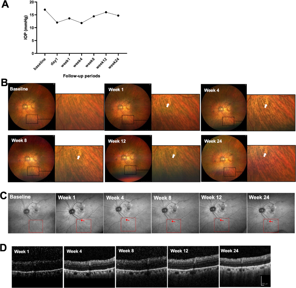
Figure 5. Safety findings for cceAAV-aflibercept-Fc-YTE (F-CRG-B191) in nAMD patient
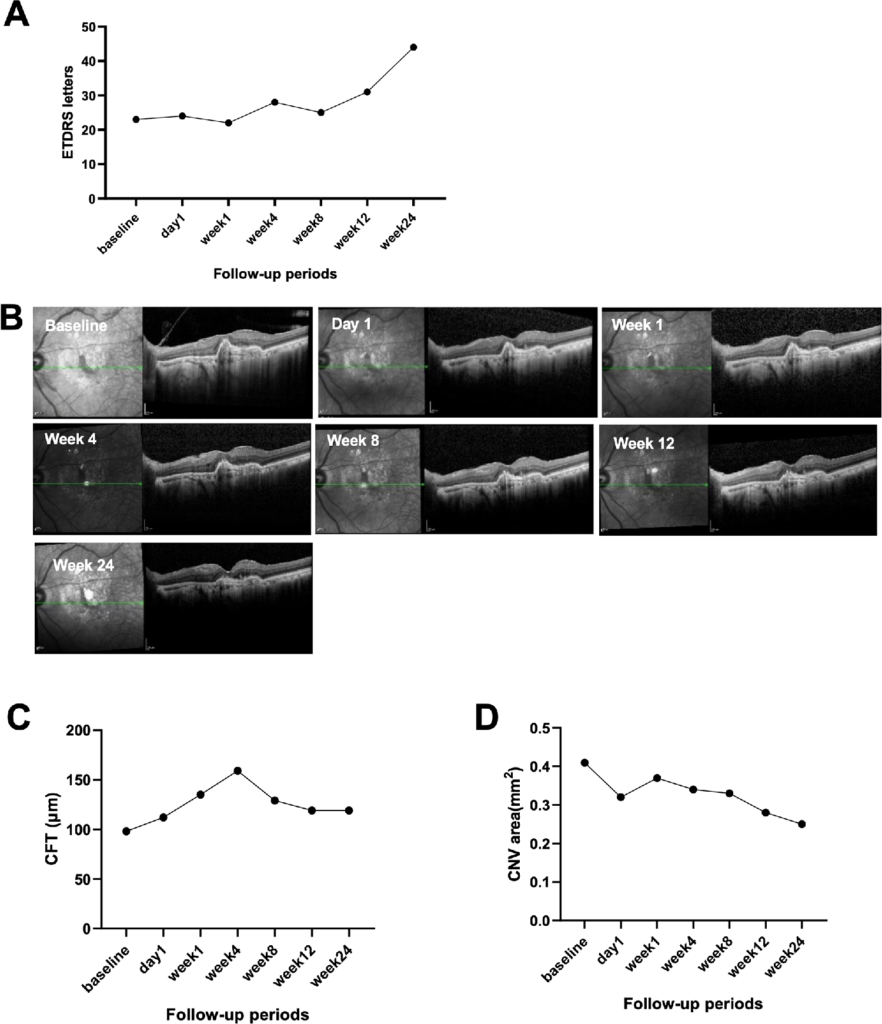
Figure 6. Efficacy evaluation of cceAAV-aflibercept-Fc-YTE (F-CRG-B191) in nAMD patient.
Conclusion
We designed and developed cceAAV-aflibercept-Fc-YTE (FCRG-B191) as a gene therapy candidate for nAMD, and other anti-VEGF purpose. In a series of experiments, cceAAV-aflibercept-Fc-YTE (F-CRG-B191) exhibited good safety and strong anti-angiogenic properties. These findings support its use as an alternative therapy for nAMD. Future studies with larger numbers of cases, longer follow-up periods, and refined dosing protocols are necessary. Also, long-term effects on RPE and photoreceptors warrant cautious dose optimization and extended observation.
Reference
Shi, J., et al., Safety and Efficacy of cceAAV-aflibercept-Fc-YTE (F-CRG-B191): A Gene Therapy for Neovascular Age-Related Macular Degeneration. Investigative Ophthalmology & Visual Science, 2026. 67(4).