
Comparative characterization of Cas12f orthologs reveals mechanistic features underlying enhanced genome editing efficiency
Apr 17, 2026
The 2026 Nature structural & molecular biology by Guan, K., et al. researches Comparative characterization of Cas12f orthologs reveals mechanistic features underlying enhanced genome editing efficiency.
Introduction
Miniature CRISPR-Cas12f nucleases are attractive candidates for therapeutic genome editing because of their compact size and compatibility with adeno-associated virus (AAV) delivery. However, editing efficiencies in mammalian cells are lower than those of larger systems. The extensive phylogenetic diversity of Cas12f suggests unexplored mechanistic variation with the potential for optimization. Here we identify and characterize a naturally occurring Cas12f ortholog discovered through metagenomics, Alistipes sp. Cas12f (Al3Cas12f), which supports robust genome editing in human cells. Through structural, biochemical and kinetic analyses, we compare Al3Cas12f to two recently described orthologs, Oscillibacter sp. Cas12f and Ruminiclostridium herbifermentans Cas12f. These orthologs present divergent architectures and regulatory features governing protospacer-adjacent motif recognition, guide RNA (gRNA) binding, dimerization and DNA cleavage. Notably, Al3Cas12f achieves deficient R-loop formation through a stable dimer interface and a naturally optimized gRNA. Leveraging these structural insights, we generate an engineered Al3Cas12f variant (RKK) that increases editing and improves activity across several tested genomic loci. By overcoming locus-dependent variability and an apparent potency threshold, this engineered compact editor seems to expand the feasibility of low-dose, AAV-compatible therapeutic genome editing. Our results elucidate mechanistic determinants of Cas12f activity and ofer a framework for engineering compact genome editors that may bear therapeutic potential.
Finding Highlights
- Present a newly identified Cas12f ortholog from Alistipes sp., Al3Cas12f, which exhibits high editing efficiency in human cells.
- Conduct kinetic analyses of all three orthologs and performed direct comparisons of structural and biochemical features.
- Enable structure-guided engineering of Al3Cas12f to enhance activity across tested targets, supporting therapeutic development of compact genome editors
Key Results and Figure Interpretatio
- Al3Cas12f is a highly active CRISPR-Cas nuclease (Fig.1).
Although divergent, some of these nucleases are related to other Cas12f nucleases when placed in a phylogenetic tree of archetypal type V nucleases (Fig. 1a). Newly identified representative sequences Cas12f-MG119-1, Cas12f-MG119-2, Cas12f-MG119-3 and Al3Cas12f share 28–52% average amino acid sequence identity with reference sequences and range between 433 and 488 aa in length (Fig. 1a). Upon inspection of the genomic regions encoding the nuclease genes, we identified the corresponding CRISPR RNAs (crRNAs) and trans-activating crRNAs (tracrRNAs). We determined that these systems recognize T-rich PAMs using an in vitro cleavage assay of an 8N PAM library and cut the TS at 20, 22 and 23 nt from the 5′ PAM sequence and the NTS at 11 and 13 nt (Fig. 1b, Supplementary Fig. 2 and Extended Data Fig. 1). We next studied the activity of Al3Cas12f in human cells. Results from a gRNA screen targeting intron 1 of the ALB gene, exon 3 of the APOA1 gene and the AAVS1 site within PPP1R12C intron 1 showed that 27 target sites displayed >10% editing, 19 sites displayed >50% editing and 10 sites displayed >90% editing across AAVS1 and APOA1 (Fig. 1c). Together, these results suggest that Al3Cas12f is highly active in its native form. To further validate the high levels of activity observed in mammalian cells, we compared the activity of Al3Cas12f relative to other CRISPR–Cas orthologs. Al3Cas12f outperformed OsCas12f across six of eight loci and exhibited significantly higher activity than LbCas12a at two sites (NLRC4 and NUDT16) (Fig. 1d,e).
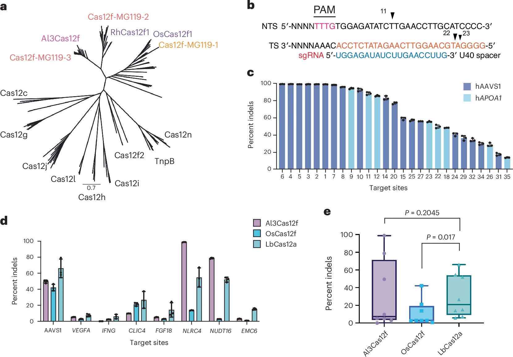
Figure 1. Initial discovery and characterization of the compact type V Al3Cas12f nuclease.
- Cryo-EM structures of Al3Cas12f, OsCas12f and RhCas12f ternary complexes reveal differences in dimer interface (Fig.2).
To understand the structural basis for enhanced activity of Al3Cas12f relative to other orthologs, we first solved a cryo-EM structure of the enzyme bound to its gRNA targeting a 55-bp target dsDNA. The Al3Cas12f structures revealed an asymmetric homodimer in a post cleavage state. The dimer is anchored by multiple interactions within its recognition (REC) domains, as well as direct base-stacking interactions with the gRNA scaffold and the R-loop. Consistent with previously solved Cas12f structures, the model revealed a bilobed ternary ribonucleoprotein (RNP) complex with the recognition lobe comprising the REC and wedge (WED) domains, while the nuclease lobe contains both RuvC and ZF domains (Fig. 2a). Al3Cas12f REC domains directly contact stem 2 of its gRNA, as opposed to CnCas12f, which has a shorter stem 2. Our structure contains a fully formed R-loop with both RuvC domains docked, as opposed to the precleavage complex observed in CnCas12f, which forms a partial R-loop (Supplementary Fig. 3). Because of these differences, we sought to obtain a better mechanistic understanding of Cas12f rearrangements as a function of R-loop formation. As there are limited Cas12f structures available, we aimed to perform a more thorough comparison of several orthologs known to have high levels of genome-editing activity. A recent publication highlighted the use of OsCas12f and RhCas12f as potential genome-editing tools. Therefore, we reasoned that obtaining structures of OsCas12f and RhCas12f bound to their respective gRNAs and target DNA might provide more insights into the mechanism of Cas12f nucleases (Fig. 2b,c, Extended Data Figs. 4 and 5 and Table 1). While all structures showed bilobed asymmetric homodimers with a fully formed 20-bp R-loop, RhCas12f and OsCas12f contained notably smaller REC domains relative to Al3Cas12f (Fig. 2b,c). In addition, only Al3Cas12f and OsCas12f had resolvable RuvC domains tethered to the R-loop, while neither RuvC domains were resolved for RhCas12f (Fig. 2c). These observations suggest that dimerization interfaces vary widely among Cas12f nucleases and that the RuvC domains can sample multiple conformational states and exhibit different levels of structural plasticity across Cas12f orthologs.
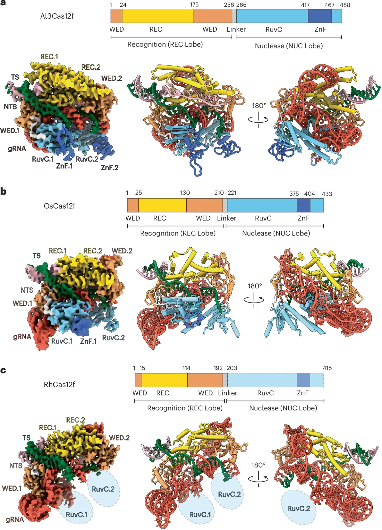
Figure 2. Cryo-EM structure of Cas12f orthologs reveal distinct dimerization interface and RuvC plasticity
- Cas12f gRNAs exhibit structural and functional diversity (Fig.3).
The Cas12f ternary complexes demonstrate notable structural diversity among the cognate gRNAs of these nucleases. The gRNA scaffolds of OsCas12f and RhCas12f share a conserved L-shaped architecture (Fig. 3a,b), like AsCas12f. The long stem 2 of the gRNA of RhCas12f and OsCas12f is nearly perpendicular relative to stems 3 and 4. While this region is not well resolved for OsCas2f, stem 2 in the RhCas12f gRNA exhibits a 90° kink that creates additional stabilizing contacts with the Cas12f monomer 2, making it highly resolvable in our structure (Fig. 3b). This region docks into the groove formed by the REC and WED domains and contacts the PAM-interacting residues of monomer 2. Notably this region of the gRNA shares the same sequence with its PAM and is recognized by the same residues involved in PAM binding by monomer 1. Specifically, a hydrogen bond with the C57 (Fig. 3c and Extended Data Fig. 7). These observations reveal that the same residues in each RhCas12f monomer facilitate dual recognition of the PAM and gRNA, respectively, and prevent nonproductive PAM binding by the wrong monomer. To examine whether the distal end of stem 2 is necessary for Cas12f activity, we deleted this region in the gRNA of OsCas12f (Δ40–58 nt) and RhCas12f (Δ50–60 nt) and assessed their activity in a plasmid interference assay. Deletion of the flexible region in stem 2 did not affect OsCas12f activity. In contrast, RhCas12f activity was significantly reduced (Fig. 3d), indicating that the interaction between Cas12f.2 and stem 2 of the gRNA is essential for RhCas12f activity. As our structures indicated that stem 4 in OsCas12f and RhCas12f did not form direct contacts with the protein, we speculated that deletion of this region could enhance the efficiency of these enzymes. Indeed, deletion of the stem 4 region (Δ106–125 nt) in OsCas12f showed a tenfold increase in plasmid interference (Fig. 3d). We conclude that Al3Cas12f contains a naturally optimized gRNA scaffold and supports previous findings that removal of stem 4 in Cas12f nucleases similarly enhances nuclease activity23. Overall, our results provide new mechanistic and structural insights into the gRNA configuration and optimization of Cas12f nucleases.
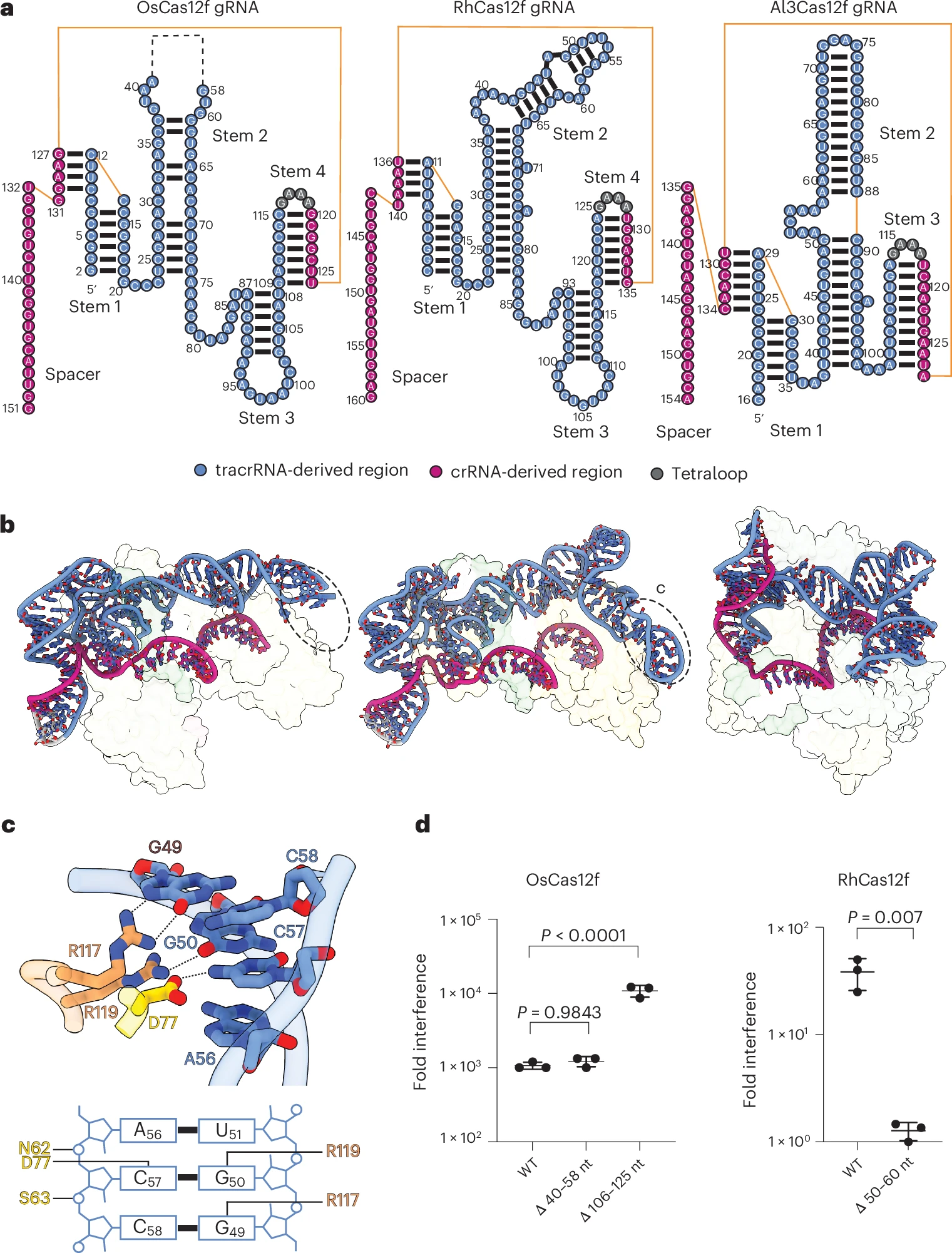
Figure 3. Overview of gRNA architectures of OsCas12f, RhCas12f and Al3Cas12f
- Variability in the Cas12f dimerization interface explains enhanced activity by Al3Cas12f. (Fig.4).
Cas12f dimerizes through interactions between the REC domains of its two monomers. OsCas12f and RhCas12f primarily form dimers through two-layer interactions; the top layer involves the first α-helix of the REC domain (α1), while the bottom layer is formed by two loops located below α1 (Fig. 4a–f). In contrast, the dimerization interface of Al3Cas12f is more complex compared to OsCas12f and RhCas12f (Fig. 4g–k). The REC domain of Al3Cas12f is larger, containing an additional 50–60 residues that form two α-helices (α2 and α3). These extended helices in REC.1 and REC.2 interact with the NTS DNA and the hairpin in stem 2 of the gRNA, respectively (Extended Data Fig. 8a–c). These REC domain interactions with nontarget DNA and gRNA have not been observed for other Cas12f nucleases and may result in enhanced stability of the complex. Al3Cas12f contains an α-helix α1 in REC.2 (α1.2) that curves toward REC.1, while α1 of REC.1 (α1.1) remains mostly linear (Fig. 4g). The bending of α1.2 toward α1.1 allows α1.1 to dock onto the α1.2 helix, aided by multiple π–π stacking interactions. This unique dimerization mechanism is analogous to a mortise-and-tenon model, where the mortise is the concave slot of the receiving part (α1.2) and the tenon is the protruding structure that fits into the mortise (α1.1) (Fig. 4g). Moreover, Al3Cas12f contains two other mortise–tenon joints located alongside the helices of the REC domain (Fig. 4h,k). The stability at the interface of Al3Cas12f is mediated through a combination of π–π stacking interactions between aromatic residues and van der Waals interactions among nonpolar amino acids (Fig. 4j). Additionally, hydrogen bonds and salt-bridge interactions have a notable role in the stabilization of the dimer (Fig. 4h,i,k).
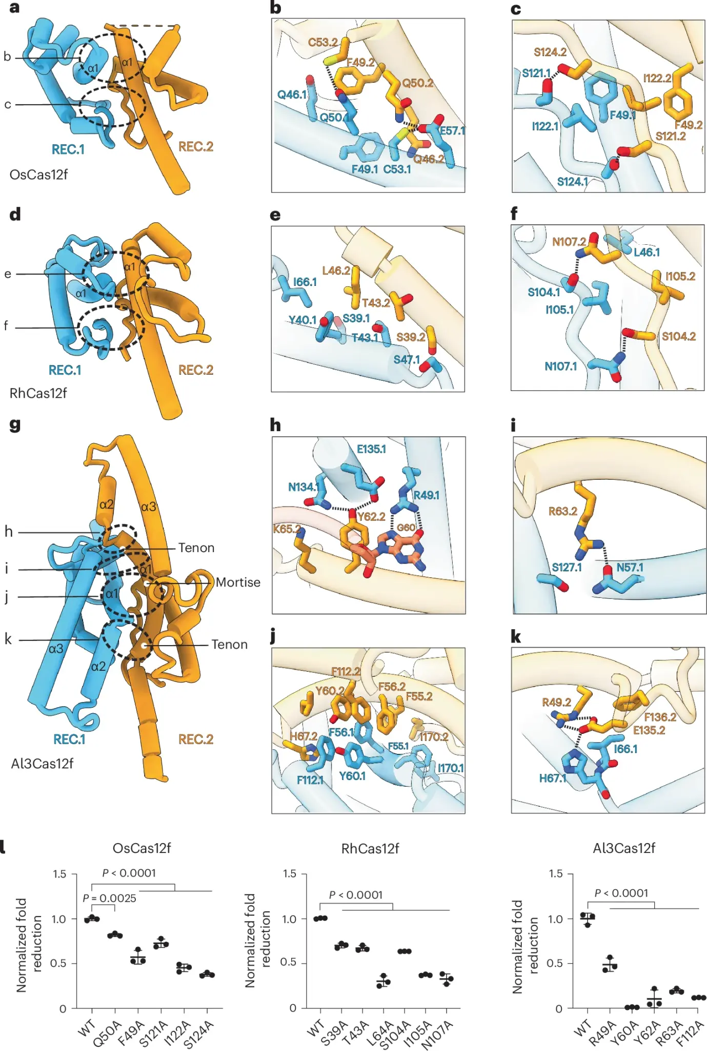
Figure 4. REC dimerization interfaces in OsCas12f, RhCas12f and Al3Cas12f
- Cas12f intermediate structures reveal differing activation mechanisms for Cas12f variants (Fig.5)
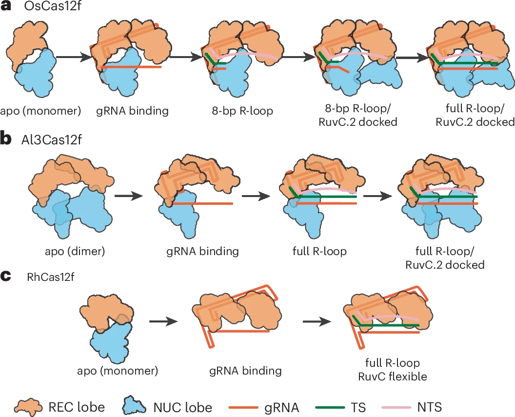
We solved three distinct structures of OsCas12f in multiple R-loop formation and conformational states (Fig. 5a–c). State I showed an 8-bp R-loop with an unresolved RuvC.2, indicating this domain can sample multiple conformational states, as previously seen for CnCas12f (Fig. 5a). State II revealed the RuvC.2 domain docked onto the spacer region of the gRNA before R-loop completion, allowing us to model six additional nucleotides of the gRNA spacer alongside the 8-bp heteroduplex, while RuvC.2 was only partially resolved (Fig. 5b). In state III, the structure displayed a fully formed 20-bp R-loop with RuvC.2 domain partially resolved (Fig. 5c). Al3Cas12f displayed two different intermediates, both with a fully formed 20-bp R-loop: a preproduct state before RuvC.2 docking (state I) and a postproduct state with a fully docked RuvC.2 domain at the PAM-distal end of the spacer (state II) (Supplementary Fig. 6). Although we show that the RuvC.1 domain in OsCas12f is nuclease active and cleaves both strands (Supplementary Fig. 7), our structures indicate that RuvC.2 undergoes notable structural rearrangements before eventually docking and stabilizing the PAM-distal region of the R-loop. This enables full R-loop formation in OsCas12f in state II. Conversely, Al3Cas12f can form a full R-loop before RuvC.2 docking. This discrepancy in the order of RuvC docking between OsCas12f and Al3Cas12f suggests mechanistic differences between these nucleases that may affect their cleavage efficiency.
To understand the importance of these intermediate structures, we examined the RuvC lid in OsCas12f and Al3Cas12f. States I and II in OsCas12f (8-bp R-loop formed) show the RuvC lid in a ‘closed’ conformation (Fig. 5d). OsCas12f State III structure is flexible and not modeled (D335–L342). We show that this lid is essential for cleavage by substituting E334–R343 to alanine, preventing the lid from undergoing the rearrangements necessary for cleavage and resulting in significantly reduced OsCas12f activity in GFP depletion assays (Fig. 5e). In addition to the RuvC loop, we noticed that W345 in OsCas12f positions DNA in the active site of the RuvC domain, like what has been observed in UnCas12f. A W345A substitution significantly hindered its activity in E. coli, indicating that this residue is essential for activity in OsCas12f (Fig. 5d,e).
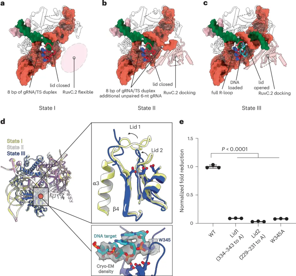
Figure 5. DNA target loading is facilitated by a two-lid opening mechanism
- Structure-guided engineering of Al3Cas12f increases editing efficiency (Fig.6)
We started by testing single substitutions and evaluating the editing efficiency of these variants. Six single-substitution variants with editing efficiency over 60% were chosen to create combinatorial mutations (Supplementary Fig. 10a). We tested 35 double-substitution and triple-substitution variants in K562 cells targeting one AAVS1 site and the SOD1 sites, as well as one hexamutant that combined six substitutions (Supplementary Fig. 10b and Fig. 6a). In total, 21 of the 35 combinatorial mutants showed at least 75% editing efficiency at SOD1 with the A9 gRNA with one quarter of the dose used in the previously described spacer length assay, 125 ng of mRNA and 50 pmol of gRNA (Fig. 6b). Only the hexamutant displayed lower levels of editing efficiency (~10%) than the wild type (~40%). From this assay the K79R;M190K;E222K variant (no. 53) was chosen as the lead Al3Cas12f variant, hereby referred to as Al3Cas12f RKK (Fig. 6a,b and Supplementary Data 1). Al3Cas12f RKK was benchmarked against the wild-type enzyme using eight reference guides previously used for AsCas12f (ref. 21). Six of these guides displayed >80% indels, with up to a 26-fold increase in activity at the EMC6 locus (Fig. 6c). Overall, these results show that Al3Cas12f RKK is a highly efficient and compact nuclease that could be further optimized for genome editing.
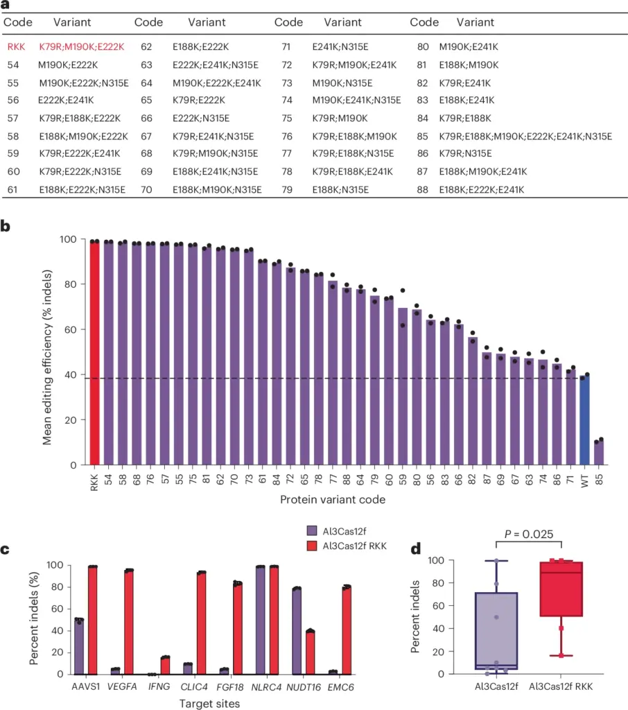
Figure 6. Structure-guided engineering of Al3Cas12f furtherly improved editing efficiency
Conclusion
Cas12f nucleases are highly desirable because of their size and levels of genome-editing activity. Through mining of a large, assembly-driven metagenomics database, we identified a natural ortholog of Cas12f, Al3Cas12f, that exhibits high levels of activity in vitro and in mammalian cells. We show that this enzyme is advantageous because of its recognition of a minimally restrictive PAM, naturally optimized gRNA and ability to remain a highly stable dimer.
Through a combination of structural, kinetic and biochemical assays, we highlighted multiple mechanistic differences among Cas12f orthologs that expand our understanding of the structural features important for achieving high nuclease activity. We showed that a highly stable dimerization interface in conjunction with a naturally optimized gRNA enables Al3Cas12f to form an efficient R-loop without requiring prior domain rearrangements. Lastly, we showed that an engineered variant increased gene-editing activity across multiple human genomic loci, making it well suited for a wide range of biotechnological and therapeutic applications that require compact nucleases for efficient delivery.
Reference
Guan, K., et al., Comparative characterization of Cas12f orthologs reveals mechanistic features underlying enhanced genome editing efficiency. Nature Structural & Molecular Biology, 2026.