
Grabody B, an IGF1 receptor-based shuttle, mediates efficient delivery of biologics across the blood-brain barrier
Apr 10, 2026
The 2022 Cell Reports Methods by Shin, J.W., et al researches Grabody B, an IGF1 receptor-based shuttle, mediates efficient delivery of biologics across the blood-brain barrier.
Introduction
To overcome the limited delivery of antibody therapeutics to the central nervous system due to the blood-brain barrier (BBB), molecular shuttles have been actively developed. The molecular shuttles were designed to bind to receptors at the surface of brain endothelial cells that are major components of the BBB. Most of the conventional molecular shuttles, however, exhibit low cross-reactivity, safety issues, or poor brain-penetrating capability. Here, we report a non-neutralizing anti-insulin-like growth factor 1 receptor (IGF1R) antibody (Grabody B) as a safe and efficient molecular shuttle that can be applicable to various therapeutic modalities for CNS-related disorders.
Finding Highlights
- IGF1R is enriched higher in the CNS than in the periphery.
- Grabody B, an anti-IGF1R antibody-based shuttle, does not cause safety issues in animals.
- Grabody B increases the BBB penetration of antibody therapeutics.
- Grabody B increases the efficacy of an anti-α-Syn antibody in a PD animal model.
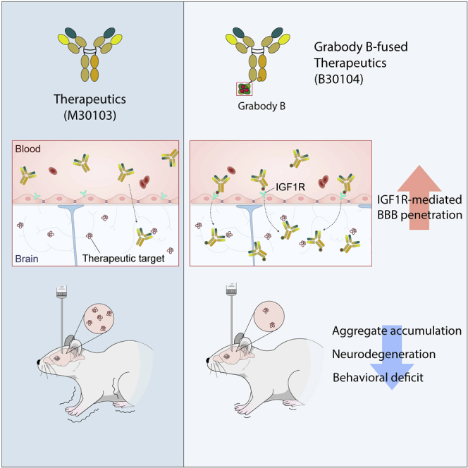
Key Results and Figure Interpretation
- IGF1R is enriched in the CNS and its vasculature (Fig.1).
To compare the IGF1R expression in the brain and peripheral organs, five mouse tissues (brain, pancreas, liver, colon, and heart) were immunoblotted with a specific antibody against the 97-kDa IGF1Rb (Figure 1A). To compare the IGF1R expression level in brain vessels and parenchyma, each fraction was separated and probed with an antibody against IGF1R. Strong IGF1R immunoreactivity was observed in both fractions, but the mean ± SEM (n=3) intensity was stronger in the vascular fraction than in the parenchyma after normalization to b-actin (Figures 1C and 1E). CD31 was highly enriched in the vascular fraction, confirming the separation of brain vessels and parenchyma (Figures 1C and 1D). These data indicate high expression of IGF1R in the brain. We tried to visualize the IGF1R expression in murine brains by immunostaining. Mouse cortex showed strong immunoreactivity to IGF1R with clear IGF1R expression on BECs in the mouse microvasculature (Figure 1H). We identified the localization of IGF1R in human cortical sections without neurodegeneration by immuno-staining with an anti-IGF1R antibody. There was clear staining in neuronal cells (Figure 1F: a, e), which was confirmed by the colocalization of IGF1R and CD31 signals (Figure 1G). This collectively suggests IGF1R expression in the brain cells and brain microvessels composed of BECs. To examine whether IGF1R is also expressed in diseased human brain samples, we imaged medial frontal cortical sections from patients with AD and age matched healthy controls after double immunostaining with anti-IGF1R and anti-CD31 antibodies (Figure 1I). The brain sections of both groups showed IGF1R (red) and CD31 (green) immunoreactivity, with fewer IGF1R and CD31 signals in the AD brain sections than in the control sections (p = 0.0283; Figure 1J). Interestingly, we observed a significant increase in colocalization between IGF1R and CD31 in the AD samples.
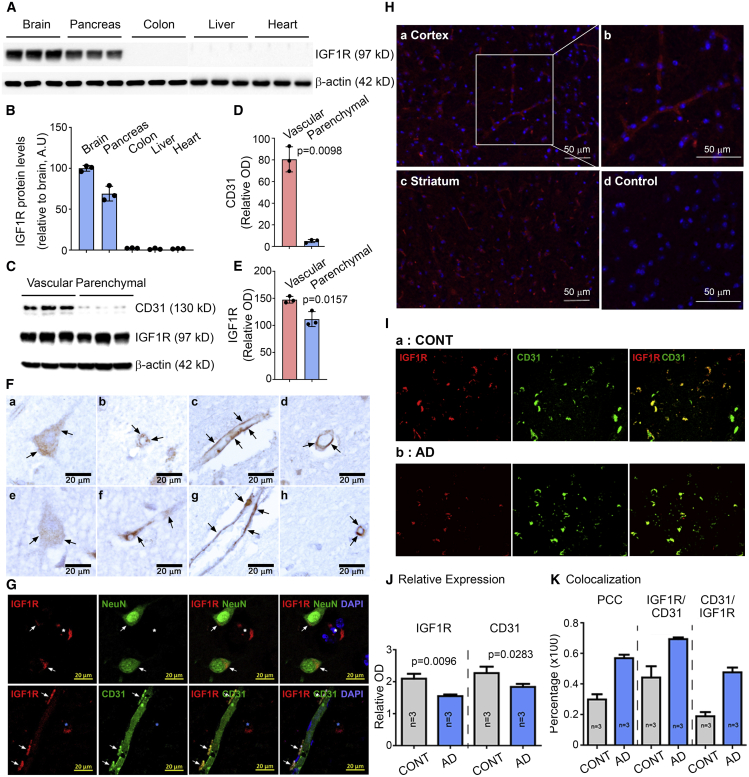
Figure 1. IGF1R is enriched in the brain and the brain vasculature.
- Grabody B does not alter IGF1R-mediated biology (Fig.2).
We pre-treated IGF1R expressing human breast cancer (MCF7) cells with human immunoglobulin G (hIgG), M30103 (anti-human a-Syn antibody; Figure 2A), B30104 (a monovalent bispecific antibody with M30103 and Grabody B, Figure 2A), or a neutralizing antibody against IGF1R (A12), followed by either vehicle or IGF1 treatment. The levels of IGF1R-mediated signaling pathway components (total AKT, phosphorylated Akt [pAKT (Ser473)], total IGF1R, and phosphorylated IGF1R [pIGF1R (Tyr1135/1136)]) were analyzed by western blot. B30104 did not affect the IGF1-induced level of pAKT or pIGF1R (Figures 2B–2D). In contrast, A12 treatment resulted in a reduction in the phosphorylation of both IGF1R and AKT. Furthermore, IGF1-induced proliferation of MCF7 cells was not affected by increasing concentrations of B30104, similar to the M30103-treated cells (Figure 2E).anti-IGF1R antibodies with neutralizing activity (MKJP2 and A12) inhibited the growth of MCF7 cells in a dose-dependent manner (Figure 2E). In line with the data above, B30104 exhibited minimal if any competition with IGF1 for interaction with IGF1R, in contrast to the dose-dependent competition of A12 with IGF1 (Figure 2F). Epitope mapping of clone #1564, a parental version of Grabody B, revealed that its IGF1R binding site is distinctively separated from the IGF1 binding sites. Taken together, these data indicate no interference in IGF1 binding or signaling through IGF1R by Grabody B.
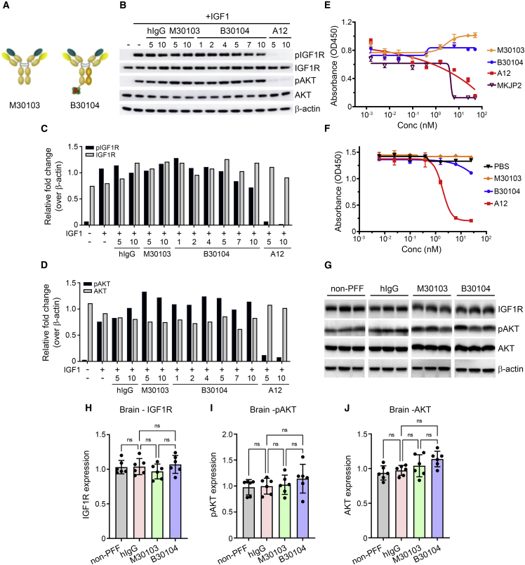
Figure 2. Grabody B does not interfere with the biological actions of IGF1R
- Grabody B increases CNS exposure to therapeutic antibodies (Fig.3).
Next, we performed intravital imaging to directly visualize Grabody B-mediated brain penetration in vivo. Seven-month-old mThy-1-a-syn transgenic mice overexpressing human a-Syn were injected with either fluorescently labeled M30103 or B30104. Alexa Fluor 555-labeled anti-CD31 antibody was co-injected to visualize brain vessels (Figure 3A). When the cortices were visualized immediately after injection and 6, 24, and 48 h post injection, the M30103 signals were mostly confined inside the brain vessels (top two rows, red, Figure 3A). In contrast, B30104 signals were redistributed from the vessels to the brain parenchyma starting at 24 h and persisted until 48 h (bottom two rows, red, Figure 3A). The differences between M30103 and B30104 with regard to their vascular versus parenchymal distributions were quantitated and presented in Figures 3B and 3C, respectively. To further track the brain localization of B30104, 4-month-old female mThy-1-a-syn mice were injected intraperitoneally with 60 mg/kg of M30103 or an equal molar amount of B30104. Mice were sacrificed at 4, 24, and 72 h. Four hours post injection, a strong green signal for B30104 was seen in brain vessels compared with M30103 (arrowhead in Figure 3D, bottom panels). At 24 h, B30104 was observed throughout the sections, including the brain vessels, brain cells, and brain parenchyma, as small puncta (Figure 3D, bottom panels). At 72 h, the signal for B30104 appeared to move further into the brain cells and parenchyma. In comparison, the M30103 signal was not detected at 4 h and was only weak in the vasculature and parenchyma at 24 and 72 h, respectively (Figure 3D, top panels). Although both antibodies were internalized into brain cells (Figure 3E), the intensity of the intracellular signal for B30104 was approximately 4-fold higher than the signal for M30103 (Figure 3F).
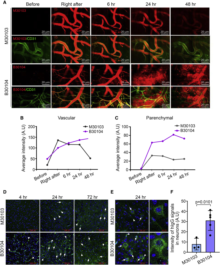
Figure 3. B30104 penetrates the brain parenchyma from brain microvessels
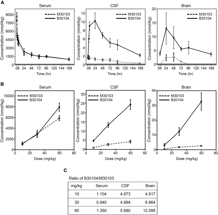
Figure 4. B30104 has sustained serum and higher sustained CNS PK profiles compared with M30103 with a wide range of BBB penetration
- Grabody B accumulates in the CNS with a significant dosing capacity. (Fig.4).
Rats were injected with 30 mg/kg of M30103 or an equal molar amount of B30104, and the serum, CSF, and brain levels of M30103 and B30104 up to 7 days were analyzed using LCSRM (Figure 4A). Importantly, when we obtained the area under the curve (AUC) of B30104 and M30103 in the CSF and brain, the AUC of B30104 was 9- to 10-fold greater than that of M30103 (Figures 4C). These data suggest that B30104 offers elevated, sustained CNS exposure up to 7 days in rodents. Next, we carried out a dosage study to determine the ability of B30104 to reach the CNS. Rats were intravenously injected with increasing doses of M30103 (10, 30, and 60 mg/kg or approximately 20.4, 60, 121.8 nM equivalent to 68, 200, 406 nmol/kg) or equal molar amounts of B30104 and their serum, CSF, and brain antibody levels analyzed by LCSRM. As shown in Figure 4B, the levels of both antibodies increased similarly in a dose-dependent manner in the blood sample. However, in both the CSF and brain, a stiff linear increase was observed with increasing doses of B30104. M30103 also demonstrated a slight increase in CNS exposure. However, even at 70.2 mg/kg (same molar amount as 60 mg/kg M30103), the level of B30104 did not appear to reach saturation in either the CSF or brain (Figure 4B). At 35.1 mg/kg (same molar amount as 30 mg/kg M30103), the brain penetration of B30104 was approximately 12-fold higher than that of M30103 (Figures 4C and S6C). Taken together, these studies suggest the efficient penetration of B30104 into the brain with relatively high saturating BBB penetration mediated by Grabody B.
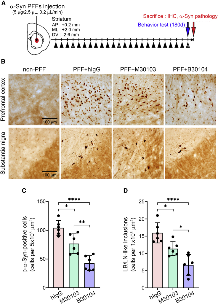
Figure 5. B30104 reduces pathological a-Syn burden more effectively than M30103
- Grabody B improves the efficacy of therapeutics for PD (Fig.5, Fig.6 and Fig.7)
We tested whether the Grabody B-mediated RMT strategy was more efficacious than an mAb in a mouse model of PD utilizing the PFF approach. Seven days post PFF injection, mice were treated weekly via intraperitoneal injection with 15 mg/kg of hIgG or M30103 or 17.55 mg/kg of B30104. At 180 days, the mice were assessed behaviorally and sacrificed for neuropathological analyses (Figure 5A). Compared with the hIgG-treated brain sections, brain sections treated with either M30103 or B30104 had a significant decrease in the number of p-a-Syn-positive cells in the prefrontal cortex and LB/LN like inclusion bodies in the substantia nigra (Figure 5B, bottom row). The quantitative results are shown in Figures 5C and 5D. Notably, B30104 was significantly more effective in reducing the brain p-a-Syn levels than M30103.
PFF injection appeared to significantly decrease the number of TH-positive neurons in the ipsilateral sections (Figure 6). This became evident in the ipsilateral side by comparing the high magnification images in the non-PFF group with the PFF-injected brains that received hIgG (Figures 6A-6C). Mice treated with M30103 or B30104 had more TH-positive dopaminergic neurons and denser tracts at 180 days post injection (dpi) than the hIgG control (Figure 6A). This was confirmed by quantification, which showed that B30104 treatment significantly increased the number of TH-positive neurons, whereas M30103 did not reach significance (Figure 6C). Overall, PFF injection eradicated approximately 50% of the TH-positive neurons at the ipsilateral SNpc, and B30104 treatment rescued approximately 80% of the ipsilateral TH-positive neurons (Figure 6D). These results collectively demonstrate that B30104 treatment effectively ameliorates neuropathological deficits in the PFF-injection model.
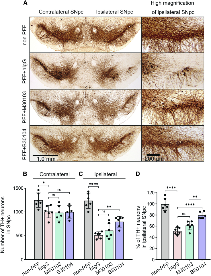
Figure 6. The therapeutic effect of RT and AAV-iIL12 is IFNγ-dependent and FAS-mediated
As shown in Figures 7A and 7B, immunoblotting the insoluble fractions from the same animals for their p-a-Syn content identified three strong, dense bands larger than 15 to 20 kDa (i.e., the size of monomeric a-Syn), at roughly 50, 75, and 100 kDa, in the hIgG-treated PFF-injected group, indicating severe oligomeric p-a-Syn burden in the PFF-injected brains. Both M30103 and B30104 significantly reduced the oligomeric band intensity compared with hIgG treatment. The non-PFF control showed minimal band intensity. We further investigated whether B30104 administration can prevent the PFF-mediated damage to the dopaminergic system using western blot analysis to determine the levels of TH and dopamine transporter (DAT). As shown in Figures 7C–7E, striatal lysates from the PFF-injected, hIgG-treated mice had a significant reduction in the level of both TH and DAT compared with the non-PFF control striatum samples. Both M30103 and B30104 increased the density of TH- and DAT-immunoreactive bands, with a significantly greater effect of B30104 for both TH and DAT. Next, we investigated whether B30104 injection also rescued behavioral deficits in the PFF mice. At both 90 and 180 dpi, mice receiving hIgG had a significant deficit in fall latency compared with the non-PFF mice in the rotarod assay. Compared with hIgG, B30104 significantly reduced the fall latency deficit at both 90 and 180 dpi, but M30103 did not (Figure 7F). The superior efficacy of B30104 in behavioral rescue was confirmed by a wire hang test to test for grip strength, which significantly reduced the fall latency deficit at both 90 and 180 dpi (Figure 7G).
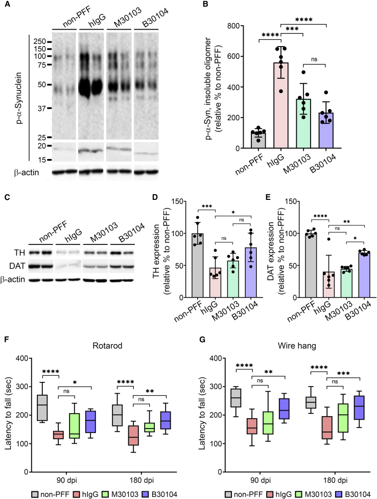
Figure 7. B30104 effectively rescues behavioral deficits by reducing pathological a-Syn and dopamine-related degeneration in the PFF injected mouse model
Conclusion
In this study, researches demonstrated that IGF1R was predominately expressed in the brain, to a lesser extent in the pancreas, but minimally in the colon, liver, and heart. We have also shown that IGF1R is expressed in both the brain vasculature and parenchyma, in both healthy and diseased human brains, and is not affected by aging.
Taken together, the results suggest that IGF1R-based Grabody B represents a new RMT platform for delivering therapeutics into the brain. Our extensive studies have demonstrated that Grabody B has superior properties for crossing the BBB without adverse effects. Unlike conventional BBB shuttles, Grabody B has broad cross-reactivity with similar IGF1R binding affinity across different species. Furthermore, Grabody B-based therapy achieves significant therapeutic outcomes in an animal model of PD. These important characteristics make Grabody B an effective and versatile RMT-based platform for delivering a wide variety of therapeutics to the brain in neurodegenerative disorders.
Reference
Shin, J.-W., et al., Grabody B, an IGF1 receptor-based shuttle, mediates efficient delivery of biologics across the blood-brain barrier. Cell Reports Methods, 2022. 2(11).