
Improved targeted delivery of antisense oligonucleotide with an antibody mask
May 29, 2026
The 2025 Nucleic Acids Research by Hsia, H.E., et al. researches Improved targeted delivery of antisense oligonucleotide with an antibody mask.
Introduction
Antisense oligonucleotides (ASOs) are synthetic nucleic acid strands designed to modulate gene ex pression by binding to RNA transcripts. In brain diseases, transferrin receptor (TfR)-targeting antibody–ASO conjugates have shown promise for brain delivery of ASOs via transcytosis in preclinical studies. This enables the more patient friendly intravenous or subcutaneous administration of the compounds. However, these conjugates can exhibit faster plasma clearance and different peripheral pharmacokinetic profiles due to ASO modifications, such as phosphorothioate (PS) linkages and locked nucleic acids (LNAs). In this study, we employed an antibody recognizing LNA- and PS-modified ASOs independent of the base sequences as a cloaking module to mitigate these issues. Using Kutzneria albida transglutaminase (KTG) technology and click chemistry, we generated TfR antibody– ASO conjugates with covalently or noncovalently incorporated ASO binders. Additionally, a noncovalent carrier antibody approach was explored. These conjugates and complexes with additional ASO binder(s) show improved TfR-targeted cellular uptake, undergo transcytosis in cellular blood–brain barrier models, show less nonspecific cellular accumulation, and anticipated ASO activity.
Finding Highlights
- Employed an antibody recognizing LNA-and PS-modified ASOs independent of the base sequences as a cloaking module to mitigate these issues.
- Use Kutzneria albida transglutaminase (KTG) technology and click chemistry, we generated TfR antibody– ASO conjugates with covalently or noncova lently incorporated ASO binders.
- Improve TfR-targeted cellular uptake, undergo transcytosis in cellular blood–brain barrier models, show less nonspecific cellular accumulation, and anticipated ASO activity.
Key Results and Figure Interpretation
- Design of < TfR/ASO > bispecific antibody ASO conjugates and complexes that enable TfR-mediated transcytosis while simultaneously “cloaking” ASOs (Fig.1).
To address nonspecific cell surface binding of antibody conjugates harboring chemically modified ASOs, we aimed at “cloaking” their ASO part with antibody-based binders and hence shield their problematic features. Figure 1 displays the three different molecular designs that we applied to explore that concept. In one approach (Fig. 1 A), antibody–ASO conjugates are complexed with 10-fold molar excess of ASO binding Fab fragments to form conjugate–Fab complexes in which the ASO is shielded by the Fab. A second approach (Fig. 1 B) is a bispecific antibody with one of its binding components being an ASO-binder to which an ASO is conjugated. This “all-in one” bispecific antibody–ASO conjugate enables intramolecular shielding of the ASO by the ASO binder on the same molecule. The third approach (Fig. 1 C) consists of a trivalent bispecific antibody harboring two ASO-binding Fab arms in close proximity, which capture ASO in a bivalent avid-binding manner to form a 1:1 antibody–ASO complex.
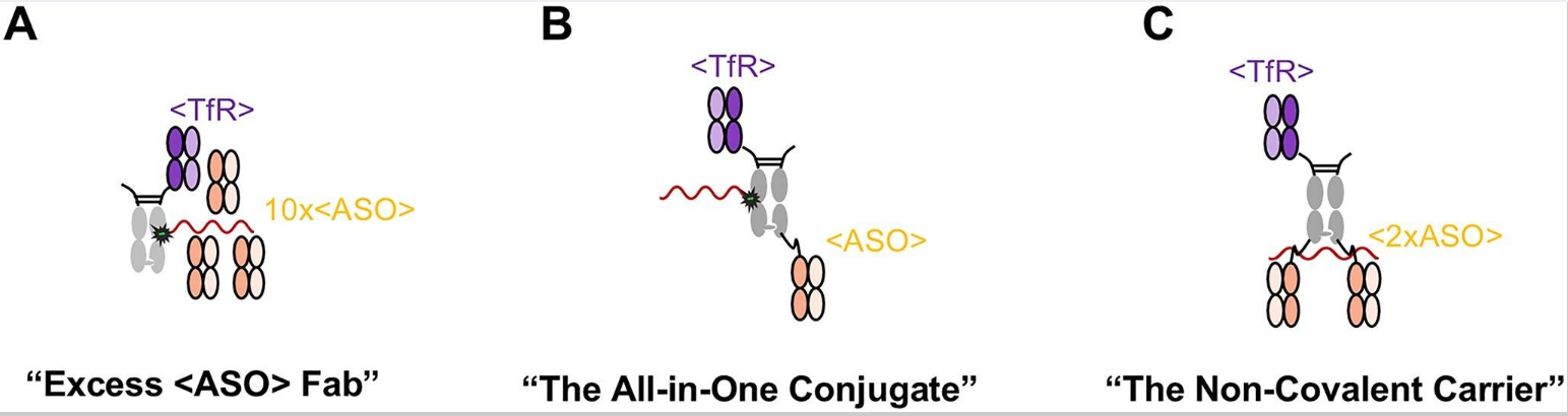
Figure 1. Three designs to include the antibody-based.
- SPR characterization of the <ASO> Fab against various LNA- and PS-modified ASOs and siRNA(Fig.2 and Table 1).
To identify a binder that is suitable to serve as a “universal” sequence-independent ASO binder defined above, we generated monoclonal antibodies by immunizing mice with modified ASOs and screened for binders that recognize modified ASOs but not unmodified nucleic acids. Three candidate binders were further characterized by assessing their specificities and binding characteristics to ASOs containing PS and LNA modifications by surface plasmon resonance (SPR). To further characterize this clone (referred to as <ASO> hereafter) while avoiding the bivalent avidity effect, <ASO> was recombinantly produced as monovalent Fab fragment. The binding of <ASO> Fab towards different PS- and LNA-modified ASOs, as well as siRNA, was tested (Fig. 2 and Table 1, left)).
Table 1. SPR results of the ASO binder (clone 1.9.21) as Fab or as IgG with two C-terminally linked ASO binder to different analytes
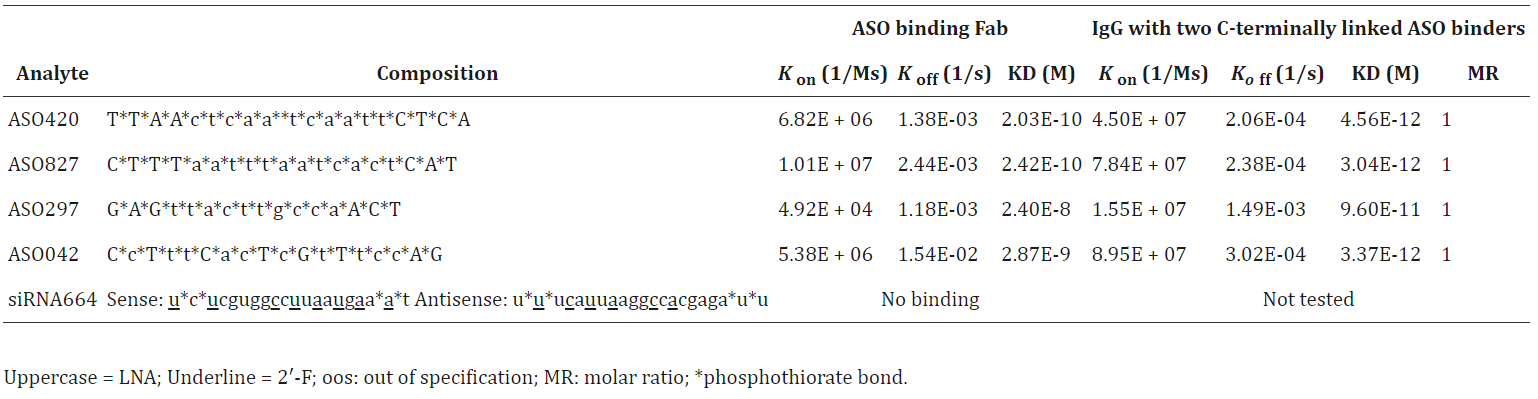
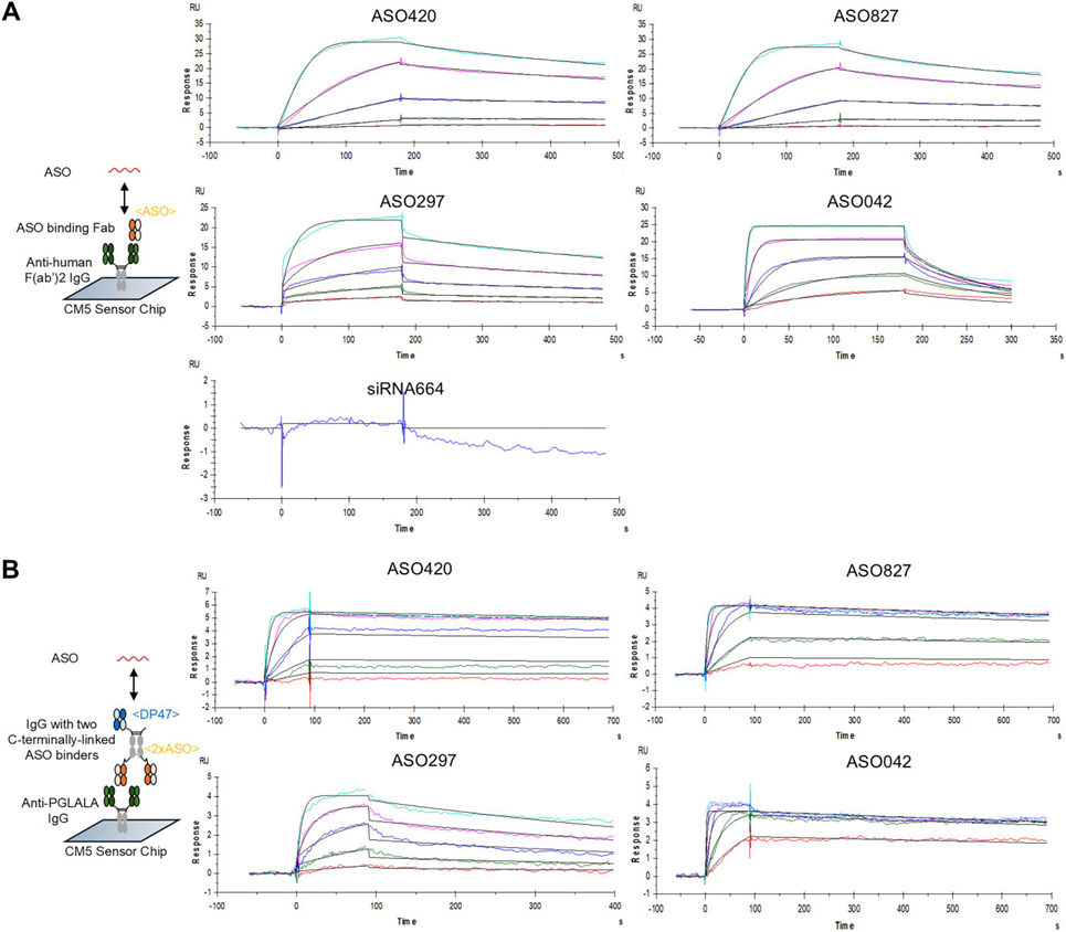
Figure 2. SPR characterization of the < ASO > Fab against various LNA- and PS-modified ASOs and siRNA.
- Structures of the <ASO> Fab in complex with short LNA- and PS- containing tetramer ASOs explain the binding specificity (Fig.3).
We determined the crystal structures of <ASO> Fab in isolation and in complex with three LNA-and PS-containing tetramers (ASO980, ASO139, and ASO143) at a resolution of 1.43-1.68 Å (Fig. 3). The binding site of the tetramer ASOs is located in the groove between the heavy and light chains, with major contributions to the paratope by the light chain involving all three complementarity-determining regions (CDRs), whereas the heavy chain mainly binds through CDR3 (Fig. 3 A–C). A hydrophobic pocket formed by the Fab accommodates the modified LNA sugar at the second position (Fig. 3 D), which correlates well with the SPR study indicating that an LNA-modified sugar is preferable at the second position (Table 2). Additional interactions include a pi–pi stacking ensemble formed by bases in positions 2–4 in combination with Tyr54 of the light chain (Fig. 3 E). Importantly, the main contacts of the tetramer ASOs with the Fab are attributed to the ASO backbone, whereas the bases are oriented outside the binding groove towards the solvent, further underscoring that the binding is base sequence-independent. Compared to ASO980 (one LNA-modified sugar), ASO139 and ASO143 have more LNA-modified sugars and form intramolecular interactions between the position 1 and 4 bases. These structural findings may explain the SPR results, where more LNA modifications correlate with higher affinity to the Fab.
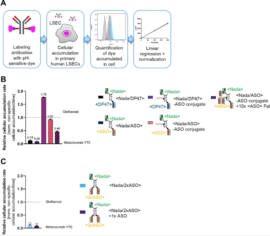
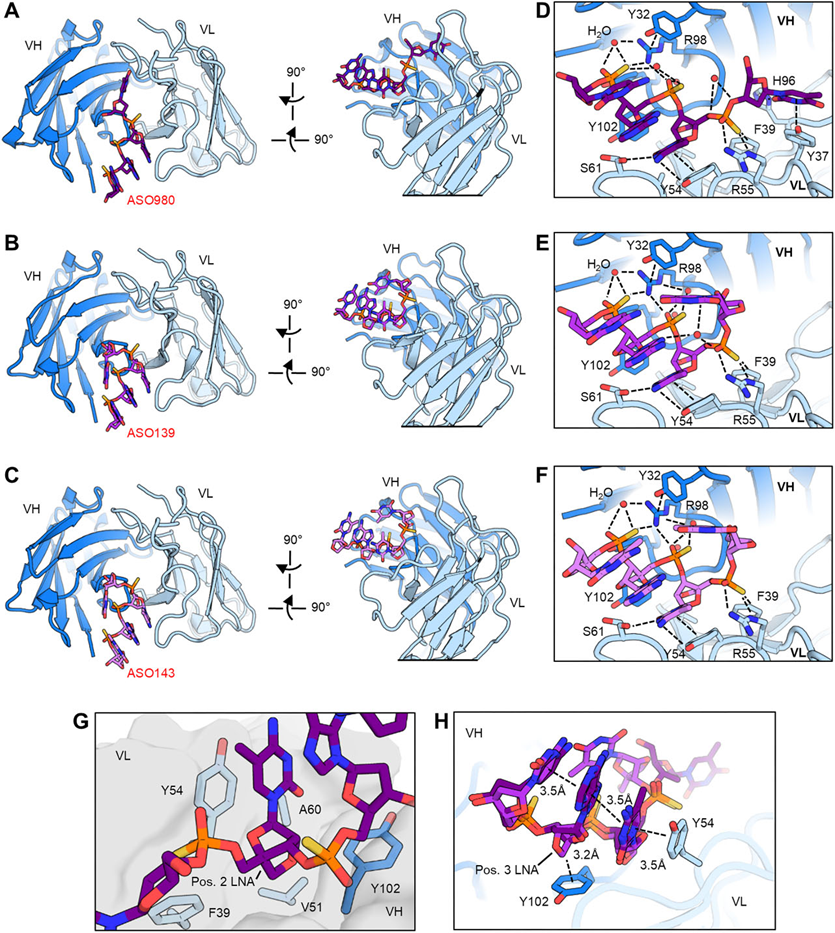
Figure 3. The <ASO> Fab engages ASOs in a sequence-independent manner via chemically modified backbones.
- The ASO binder improves transferrin receptor-dependent uptake of ASOs in hCMEC/D3 (Fig.4).
We incorporate the <AS> as a masking module to shield the ASO alongside the <TfR> for target specificity. This indicates that the ASO payload drives TfR-independent uptake of <TfR> –ASO conjugates in hCMEC / D3 cells (Fig. 4 A and B, first two conditions). The reduction in TfR colocalization of the <TfR> – ASO conjugate can be rescued by pre-incubating the conjugate with a 10-fold molar excess of the <ASO> Fab but not with the < DP47 > nonbinder as a control (Fig. 4 A and B, third and fourth columns). We also included a C-terminally linked <ASO> to the <TfR> –ASO conjugate. This approach leverages the high avidity of the bivalent <ASO> binders, which can bind a single 16–20-mer ASO with very high affinity, thereby eliminating the need to conjugate the ASO directly to the antibody. As shown in Fig. 4 C and D, ASO alone (with a 3-Atto647N fluorescence tag) can efficiently enter cells via gymnosis independent of TfR. Pre-incubation of the ASO with a 10-fold molar excess of <ASO> Fab, or an equimolar amount of a <Nada/2 × ASO> antibody (< Nada > being a nonbinder without known cell surface specificity), significantly reduced the ASO signal in cells. This indicates that the < ASO > can block ASO gymnosis.
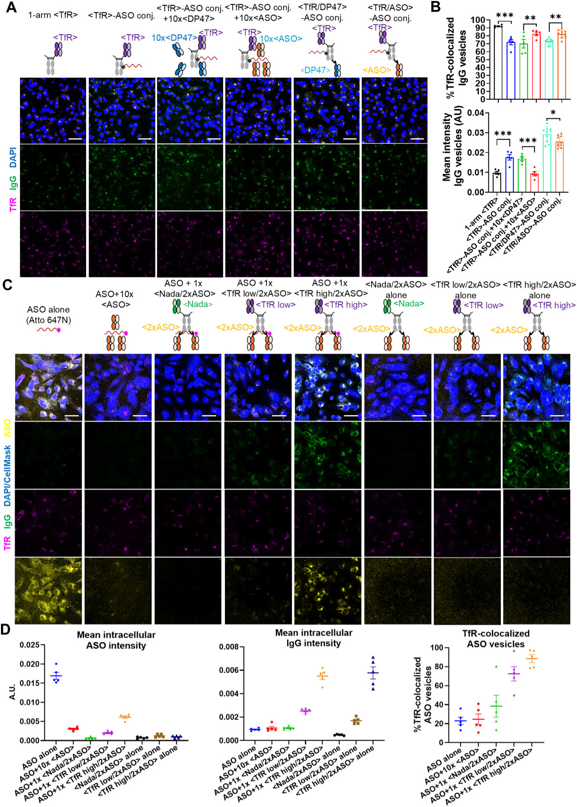
Figure 4. The ASO binder improves TfR targeted uptake of ASO in hCMEC/D3 cells
- The ASO binder decreases the transferrin receptor-independent transcytosis of ASO in spheroids (Fig.5).
After a 3-h incubation of antibodies or conjugates with the spheroid, IgG signals detected at the core of spheroids correlate with transcytosis efficiency. Results from the three different approaches to include the <ASO> are summarized in Fig. 5. In the spheroid transcytosis assay, signals from one-arm <TfR> IgG were higher at the core of spheroid compared to the negative controls, such as untreated spheroids or those treated with < DP47> control IgG, demonstrating the adequacy of this model in mimicking transcytosis (Fig. 5 A). The “all-in-one” conjugate with a C-terminally linked <ASO> was evaluated in wild-type and TfR knockout spheroids to elucidate the roles of TfR-dependent and -independent transcytosis, and to assess the impact of the additional < ASO > (Fig. 5 B). IgG with a monovalent < TfR > demonstrated efficient transcytosis in wild-type spheroids compared to the < DP47 > control, but not in TfR knock-out spheroids, indicating that IgG without the ASO payload primarily transcytoses into spheroids in a TfR-dependent manner. Importantly, conjugates with a C-terminally linked < ASO > exhibited reduced transcytosis in both wild-type and TfR knock-out spheroids compared to the < DP47 > control (Fig. 5 B). Taken together, this indicates that the additional < ASO > binder reduces nonspecific transcytosis driven by the ASO payload.
Next, we investigated the transcytosis efficiency of ASO carried noncovalently by IgG with two C-terminally linked < ASO > in spheroids (Fig. 5 C). While ASO alone (with a florescence tag) was able to transcytoses into spheroids, pre-incubating the ASO with a 10-fold molar excess of < ASO > Fab reduced the ASO signals in spheroids. Similarly, pre-incubating ASO with an equimolar amount of a < Nada / 2 × ASO > antibody, which has two C-terminally linked < ASO > but lacks cell surface specificity (< Nada >), limited the access of both ASO and IgG into spheroids (Fig. 5 C). However, pre-incubating the ASO with an equimolar amount of a “noncovalent carrier” antibody with an N-terminal low-affinity TfR binder (< TfR low / 2 × ASO >) or a high-affinity TfR binder (< TfR high / 2 × ASO >) enabled the transcytosis of both IgG and ASO into spheroids.
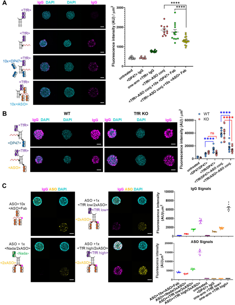
Figure 5. The ASO binder reduces TfR-independent transcytosis of ASO in BBB spheroids
- The ASO binder reduces nonspecific cellular accumulation driven by the ASO in primary human liver sinusoidal endothelial cells (Fig.6).
we determined their cellular accumulation rate in primary human liver sinusoidal endothelial cells (LSECs) as these cells are highly involved in the nonspecific clearance processes of antibodies (Fig. 6 A). Briefly, antibodies or conjugates were labeled with a pH sensitive fluorescent dye that exhibits high fluorescence when accumulated in the late endosome or lysosome (acidic pH 4– 5), but low to no fluorescence when residing outside the cell (neutral pH 7.4). Labeled antibodies or conjugates were incubated with primary LSECs for 2 and 4 h, and the fluorescent readout was recorded using a flow cytometer. The geometric mean intensities were used for linear regression analysis after subtraction of background signals (cellular auto-fluorescence) and normalization to the fluorescence of the dosing solution to account for differences in dye-to-antibody ratio. The extracted slopes indicate the cellular accumulation rate after normalization to internal standards (Motavizumab YTE= 0; Glofitamab =1) to account for experiment-to-experiment variability. As shown in Fig. 6B, antibodies without known cell surface specificities (first two columns: < Nada / DP47 > and < Nada / ASO > ) had low cellular accumulation rate, indicating low nonspecific clearance. Importantly, the “noncovalent carrier” antibody with an N-terminal < Nada > and two C-terminally linked < ASO > ( < Nada / 2 × ASO > ), with or without pre-incubation with an equimolar amount of free ASO, was also tested in these cells. The addition of free ASO to the construct did not increase the cellular accumulation rate, indicating that the ASO is cloaked by the two ASO binders (Fig. 6 C).
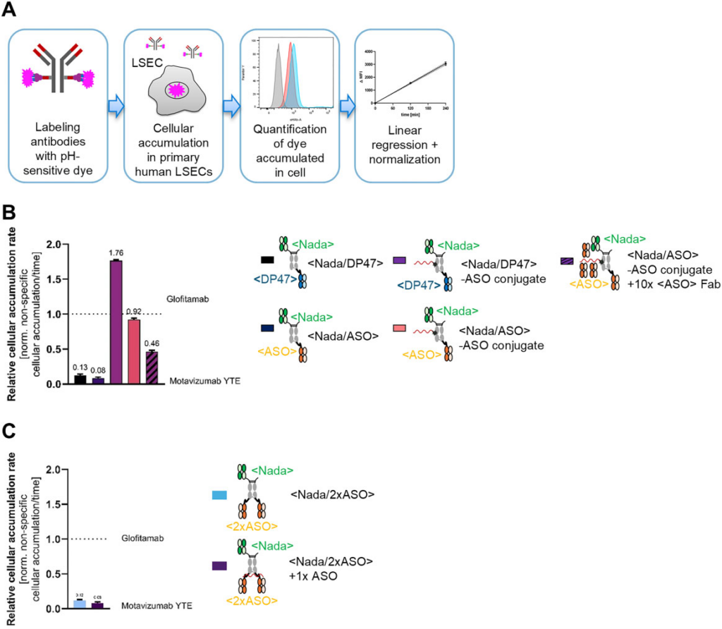
Figure 6. The < ASO > reduces nonspecific cellular accumulation driven by the ASO in primary human LSECs
- Bispecific <TfR/ASO>–ASO conjugates retain target knockdown activity in human-induced pluripotent stem cell (iPSC)-derived neurons (Fig.7).
Compounds containing ASO420 were administered four times to iPSC-derived neurons before harvesting and tested for MAPT knockdown via qPCR (Fig. 7 A). All constructs, except the non-Tau targeting control ASO, led to more than 25% MAPT knock-down, with naked Tau-targeting ASO exhibiting the highest activity (minimal fit value = 5.92%) This finding confirms previously published results and supports our data in hCMEC / D3 cells suggesting that gymnosis is more efficient for ASO uptake compared to TfR-mediated uptake. In a separate set of experiments, we also tested the activity of ASO shuttled by the “noncovalent carrier” with two C-terminally linked < ASO > (Fig. 7 B).
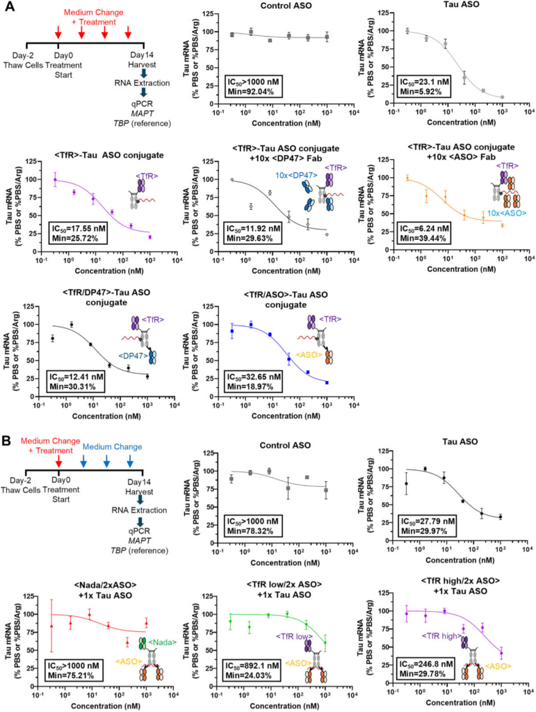
Figure 7. The <ASO> does not abolish ASO activity in human iPSC-derived neurons.
- The ASO binder improves ASO plasma pharmacokinetic profile and increases ASO brain exposure in vivo in mice (Fig.8).
Mice were administered a single IV dose of compounds containing ASO at a molar equivalent to 0.9 mg / kg of naked ASOs, ensuring uniform ASO dosing across all groups (Fig. 8 A). The animals were sacrificed after 168 h. To prevent blood contamination of ASOs in brain tissues, the animals were perfused before termination. The plasma PK profile for the naked ASO exhibited a typical pattern with a rapid distribution phase followed by a slow elimination phase (Fig. 8 B). ASO pre-incubated with the < Nada / 2xASO > antibody demonstrated very low clearance over 168 h, indicating minimal tissue distribution and effective recycling with the antibody, resulting in a prolonged plasma half-life. Regarding brain tissue distribution (Fig. 8 C), naked ASO or ASO in complex with the <Nada/2xASO> antibody did not distribute to the brain as expected, whereas the ASO conjugated or preincubated with antibodies containing a <mTfR> resulted in concentrations ranging from 18 to 32 nM, with the highest concentration observed with ASO noncovalently complexed with the <mTfR /2xASO> antibody. Taken together, the <ASO> helps extend ASO plasma PK and increases brain exposure when combined with the TfR binder in vivo.
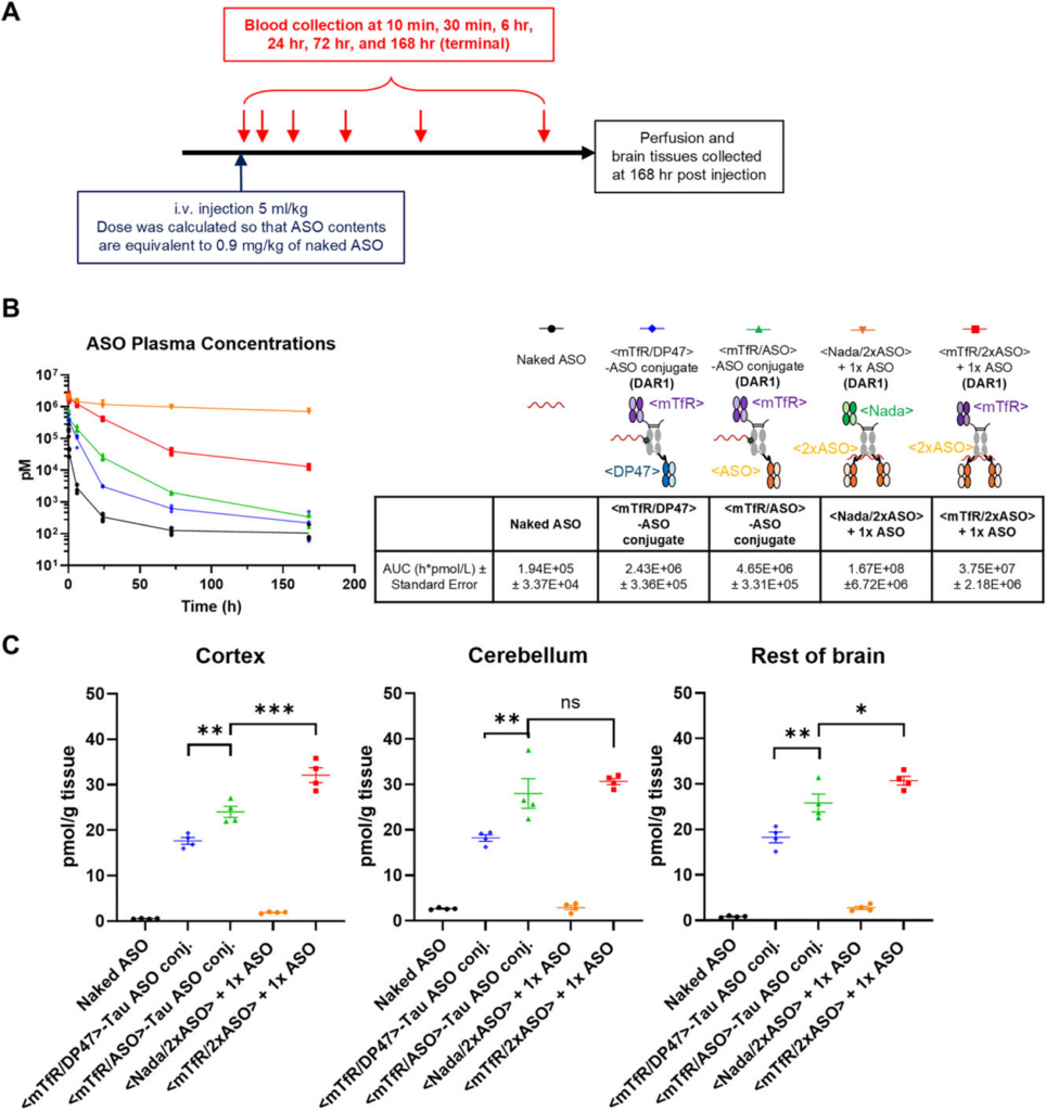
Figure 8. The <ASO> improves ASO plasma PK profile and increases ASO brain exposure in vivo in mice.
Conclusion
The addition of an ASO-cloaking binder reduces the peripheral clearance of antibody–ASO conjugates and can thus be a viable strategy, even though the resulting entities are more complex than conventional conjugates. Although including the ASO binder as separate Fab fragments to the antibody–ASO conjugate sometimes showed better in vitro properties compared to the “all-in-one” conjugate with a covalently linked ASO binder, additional Fabs require separate production and analytics and the conjugation to <TfR> requires excess ASO. Considering that ASO is costly, a “noncovalent carrier” approach using two ASO binders to hold one ASO molecule is an attractive alternative, as it does not require excess ASO.
However, whether and how the noncovalent carrier approach fits regulatory requirements for therapeutic drugs still needs to be defined. Future studies may focus on confirming the in vivo activity of ASO delivered via the noncovalent carrier approach and addressing regulatory considerations for their clinical applications.
Reference
Hsia, H.E., et al., Improved targeted delivery of antisense oligonucleotide with an antibody mask. Nucleic Acids Res, 2025. 53(20).