
Systemic delivery of AAV5, AAV8, and AAV9 packaging a C5-12-microdystrophin-FLAG expression cassette in non-human primates
June 12, 2026
The 2025 Mol Ther Methods Clin Dev by Liu, M., et al. researches Systemic delivery of AAV5, AAV8, and AAV9 packaging a C5-12-microdystrophin-FLAG expression cassette in non-human primates.
Introduction
Safely achieving therapeutic expression levels with adeno-associated virus (AAV) gene therapy is a significant challenge for treating the large muscle mass in humans. Non-human primates (NHPs) provide a more accurate assessment of the feasibility of achieving an effective and safe dose than rodents. Here, we compared a single systemic administration of AAV5, AAV8, or AAV9 in NHPs, each packaging the C5-12-microdystrophin-FLAG expression cassette. At 1month post-dose, we compared tissue vector genomes, mRNA, and microdystrophin-FLAG protein levels by meso-scale discovery-enzyme linked immunosorbent assay, liquid chromatography-mass spectrometry, and immunofluorescence. The C5-12 promoter was highly selective for heart and skeletal muscles, when compared to off-target tissues such as peripheral blood mononuclear cells, lung, liver, and kidney. AAV8 led to higher levels of microdystrophin-FLAG mRNA and protein in the cardiac ventricles and skeletal muscles when compared to AAV5 or AAV9. The AAV8-microdystrophin-FLAG led to 20% of wild-type NHP dystrophin protein expression levels and was located on the sarcolemma of 40% of skeletal muscles fibers and 15% of left ventricular cardiomyocytes. Hematology, serum chemistry, and pathology were unremarkable. Thus, a systemic dose of ~1.18×10¹⁴ vector genomes/kg AAV8 is predicted to be safe and efficacious for treating Duchenne muscular dystrophy (DMD) but has significant room for improvement.
Finding Highlights
- Assess the tropism of AAV5, AAV8, and AAV9 in in the cynomolgus monkey (Macaca fascicularis) via baculovirus expression vector system (BEVS).
- Found that each serotype effectively transduced striated muscle, and the C5-12 promoter was highly selective for both cardiac and skeletal muscles in NHPs.
Key Results and Figure Interpretation
- Selecting NHPs with low levels of nAbs (Fig.1).
Pre-treatment serum samples from 15 NHPs were screened by a cellbased nAb assay and an enzyme-linked immunosorbent assay (ELISA)-based total antibody assay (Tables 1 and S1). Five animals were excluded based on high levels of pretreatment antibodies and therefore had no group assigned to them. This resulted in 10 NHPs available for the study. Of these, 8 NHPs were identified to have low nAbs for all three tested AAV serotypes (Table 1). Post-treatment serum samples from days 15 and 29 were analyzed in the cell-based nAb assay to determine the level of nAbs against AAV5, AAV8, and AAV9 serotypes. The post-treatment nAbs were specific to the administered serotype with no cross-reactivity, with only one exception being treatment group ID 2102, which was administered AAV8 but developed nAbs to AAV8 and AAV9. Interestingly, the AAV5 samples that were neutralized by AAV8 and/or AAV9 in pre-treatment (group IDs 1102 and 1103) were no longer neutralized by AAV8 or AAV9 post-treatment.
To estimate the clearance of each AAV-C5-12-microdystrophinFLAG, plasma concentrations of the AAV VG were measured up to 48 h after treatment (Figure 1). AAV5 was mostly cleared from the blood within 24 h, while AAV8 and AAV9 remained for a period longer than 48 h (Figure 1).
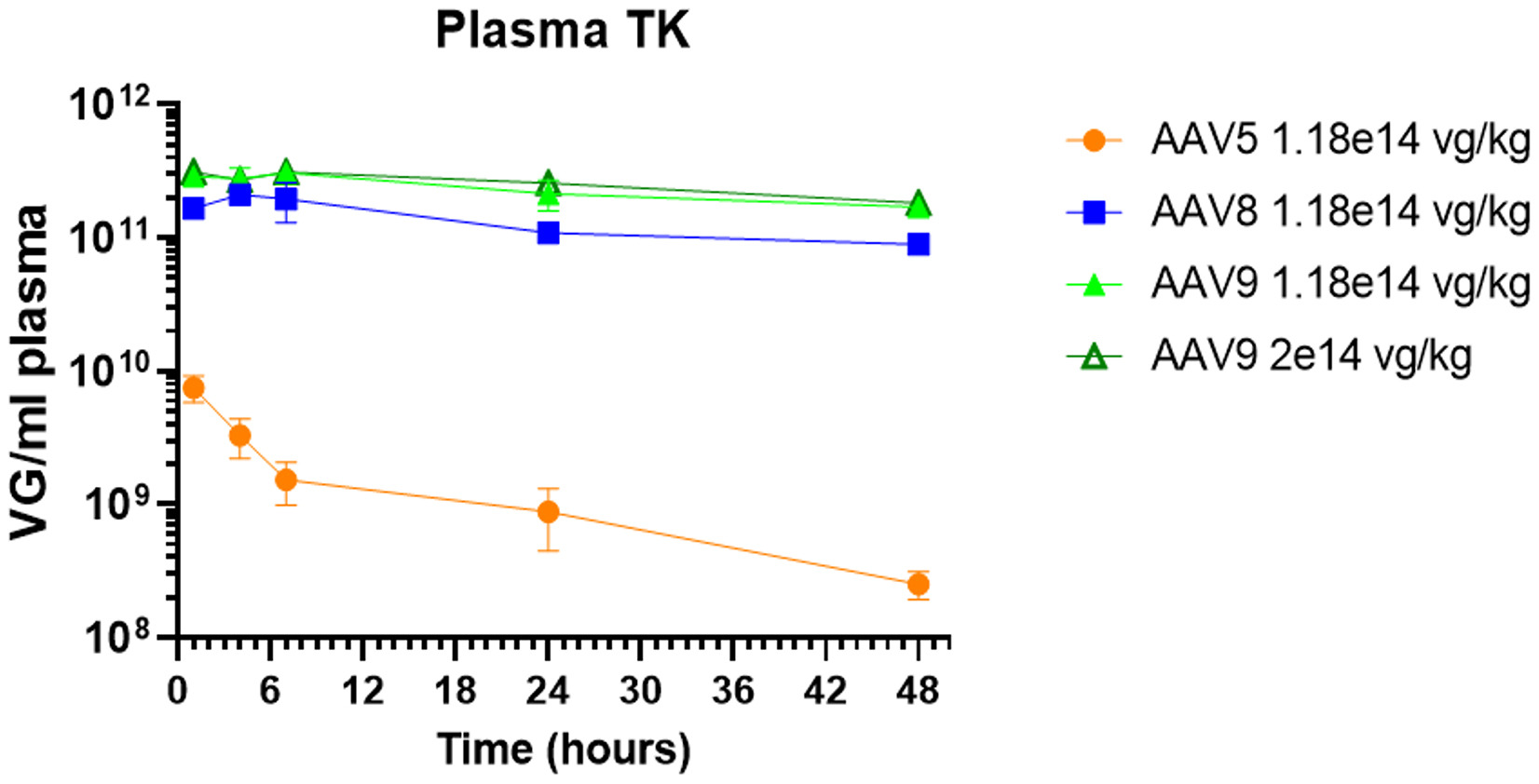
Figure 1. Plasma toxicokinetics (TK).
- Biodistribution and expression in the hear (Fig.2).
We next measured the biodistribution and microdystrophin-FLAG expression in the heart. The highest VG levels were in the atria, but this translated to minimal levels of mRNA and no microdystrophin-FLAG protein in the left atria and low levels in the right atria (Figure 2). In other regions of the heart, a narrow range of 1×10⁵ to 4×10⁵ VG/mg genomic DNA (gDNA) led to a similarly narrow range between 1×10⁵ and 6×10⁵ mRNA copies/mg total RNA (Figure 2A). The mRNA translated to low levels of protein with no clear difference in translation between the tested serotypes (Figure 2B). However, AAV5 and AAV8 led to the highest expression in the left and right ventricles when compared to AAV9 at the same dose of 1.18 x10¹⁴ VG/kg (Figures 2C and S1). Each AAV serotype had the highest expression in the ventricles, septum, and apex when compared to the atria (Figure 1C)
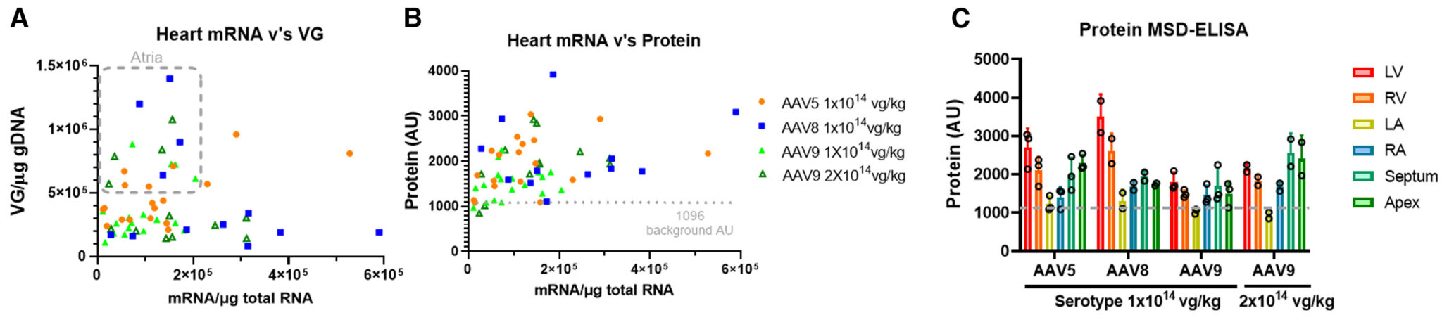
Figure 2. Expression and biodistribution in the heart after systemic delivery of AAV-C5-12-micordystrophin-FLAG
- Biodistribution and expression in the skeletal muscles (Fig.3).
The expression profile was different in skeletal muscles when compared to the heart. A narrow range between 1 and 2×10⁵ VG/mg gDNA led to 10-fold higher levels of mRNA/mg total RNA in skeletal muscles when compared to the heart (Figures 3A and S2; unfortunately, we did not measure the AAV9-treated diaphragm samples). mRNA levels below 1×10⁶ mRNA/mg total RNA led to low levels of microdystrophin-FLAG protein similar to that of the heart (Figure 3B). However, levels above 1×10⁶ mRNA/mg total RNA led to 3- to 4-fold higher levels of microdystrophin-FLAG protein (Figure 3B). One of the NHPs treated with AAV8 (group ID 2102) had the highest levels of protein in the diaphragm, quadriceps, gastrocnemius, forearm, and tongue, but not mandible or esophagus (Figure 3C). Interestingly, this NHP also developed cross-reactive levels of nAbs to AAV9 (Table 1).
We also examined microdystrophin-FLAG protein expression by liquid chromatography-mass spectrometry (LC-MS) to measure the proportion of the endogenous full-length dystrophin levels. We compared the highest dose of AAV9 (2×10¹⁴ VG/kg) to AAV8 at 1.18×10¹⁴ VG/kg (Figure 3D). The results are consistent with AAV8 leading to more protein expression than AAV9, and these levels are approximately 20% of wild-type dystrophin levels with a dose of 1.18×10¹⁴ VG/kg AAV8 (Figure 3D).
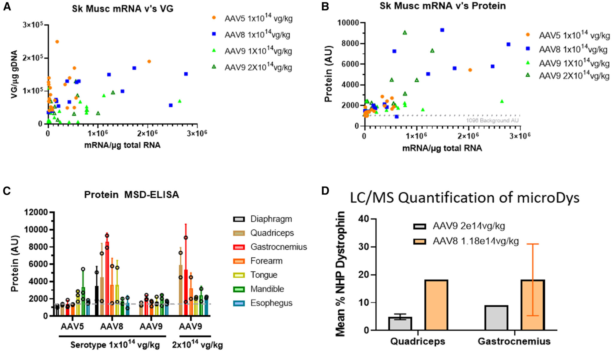
Figure 3. Expression and biodistribution in the skeletal muscles after systemic delivery of AAV-C5-12-micordystrophin-FLAG
- Proportion of striated muscles expressing microdystrophin-FLAG (Fig.4).
The proportions of cardiomyocytes and skeletal muscle fibers expressing microdystrophin-FLAG were examined by confocal microscopy with an automated algorithm to define a positive cell. Both a2-laminin and endogenous dystrophin immunofluorescence were used to mark the positive muscle membranes (Figure S3). Consistent with both the meso-scale discovery (MSD)-ELISA and LC-MS data, the AAV8 (1.18e14 VG/kg) led to the broadest expression of microdystrophin-FLAG when compared to the higher dose (2e14 VG/kg) of AAV9 (Figure 4).
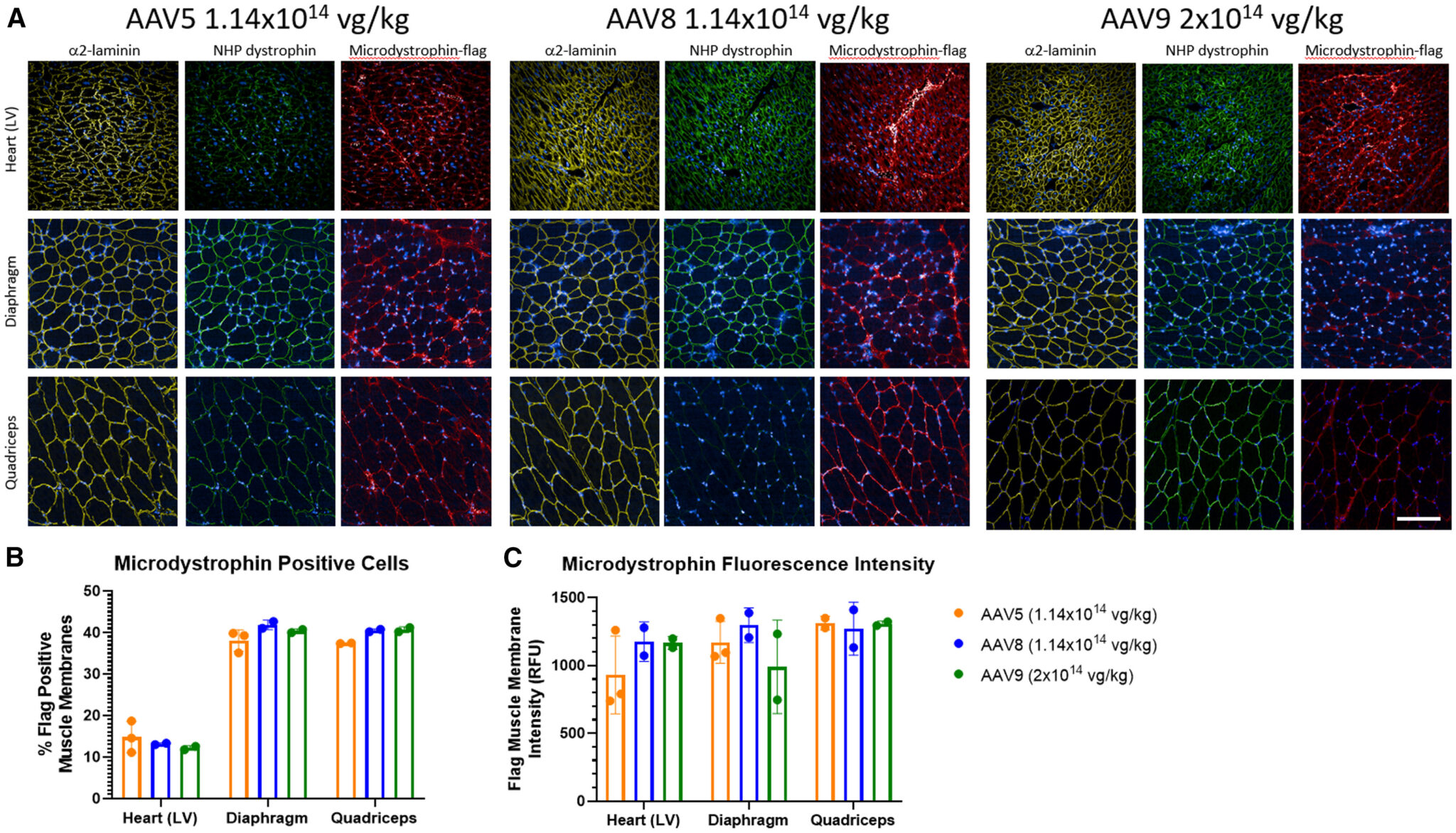
Figure 4. Microdystrophin-FLAG cellular distribution.
- Off-target tissues (Fig.5).
As anticipated, the highest levels of VG were found in the liver (Figure 5A). The VG liver, lung, and peripheral blood mononuclear cells led to background levels of microdystrophin-FLAG protein (1,096 a.u.; Figures 5B and 5C). Only the kidney for AAV5 and AAV9 treatment and liver for AAV9 treatment had microdystrophin-FLAG protein levels marginally above baseline (Figure 5), showing the excellent selectivity of the C5-12 promoter for striated muscles.
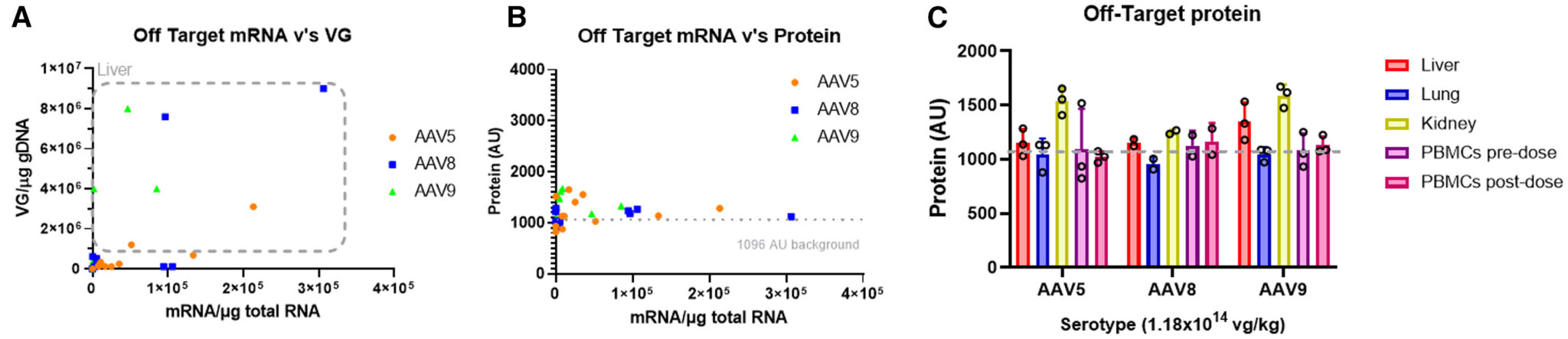
Figure 5. Expression in off-target tissues.
Conclusion
Overall, systemic delivery of AAV8 led to better biodistribution and protein expression in striated muscles of NHPs when compared to AAV5 and AAV9. The VG in the tissues were similar between serotypes, and this may have resulted from the short timeline of this study (1 month) as it may require longer time periods to clear VG that did not effectively form stable concatemers in the nucleus. Comparisons of VG, mRNA, and protein levels clearly reveal that VG delivered by AAV8 led to more effective transcription of microdystrophin-FLAG mRNA, and this greater level of mRNA led to more muscle cells expressing more microdystrophin-FLAG protein. AAV8 is also superior to AAV9 in neonatal dog hearts. The biology behind this is unclear. Perhaps one explanation is that AAV8 protects the VG from TLR9-mediated immune responses better than either AAV5 or AAV9, but this would need to be tested.
Each cardiac and skeletal muscle disease will have its own expression requirements to achieve efficacy. For DMD, approximately 20% of mini-dystrophin protein levels lead to substantial efficacy in transgenic mice when the protein is expressed in every cell. Here, delivery of 1.18×10¹⁴ VG/kg of AAV8-microdystrophin-FLAG led to 20% wild-type dystrophin protein levels in approximately 30%–50% of skeletal muscle fibers and 17% of left ventricle cardiac cells. Pre-clinical and clinical studies predict that expression as low as 5% can lead to efficacy and that 15% of normal levels of dystrophin in all muscle cells can prevent dystrophic pathology, depending on the functionality of the truncated dystrophin. More localized delivery methods such as intramuscular injection and isolated limb perfusion can achieve much greater levels of expression, but these localized delivery methods are not ideal for treating the vast muscle mass in humans. This study also serves as the basis for comparing capsid evolution efforts, which are already showing great promise in reaching more striated muscles with lower doses in NHPs.
Reference
Liu, M., et al., Systemic delivery of AAV5, AAV8, and AAV9 packaging a C5-12-microdystrophin-FLAG expression cassette in non-human primates. Mol Ther Methods Clin Dev, 2025. 33(1): p. 101411.